Smad3 Antibody - #AF6362

| Product: | Smad3 Antibody |
| Catalog: | AF6362 |
| Description: | Rabbit polyclonal antibody to Smad3 |
| Application: | WB IHC IF/ICC |
| Reactivity: | Human, Mouse, Rat |
| Prediction: | Pig, Bovine, Horse, Sheep, Rabbit, Dog, Chicken, Xenopus |
| Mol.Wt.: | 58kDa; 48kD(Calculated). |
| Uniprot: | P84022 |
| RRID: | AB_2835210 |
Product Info
*The optimal dilutions should be determined by the end user.
*Tips:
WB: For western blot detection of denatured protein samples. IHC: For immunohistochemical detection of paraffin sections (IHC-p) or frozen sections (IHC-f) of tissue samples. IF/ICC: For immunofluorescence detection of cell samples. ELISA(peptide): For ELISA detection of antigenic peptide.
Cite Format: Affinity Biosciences Cat# AF6362, RRID:AB_2835210.
Fold/Unfold
DKFZP586N0721; DKFZp686J10186; hMAD 3; hMAD-3; hSMAD3; HSPC193; HST17436; JV15 2; JV15-2; JV152; LDS1C; LDS3; MAD (mothers against decapentaplegic Drosophila) homolog 3; MAD homolog 3; Mad homolog JV15 2; Mad protein homolog; MAD, mothers against decapentaplegic homolog 3; Mad3; MADH 3; MADH3; MGC60396; Mothers against decapentaplegic homolog 3; Mothers against DPP homolog 3; SMA and MAD related protein 3; SMAD 3; SMAD; SMAD family member 3; SMAD, mothers against DPP homolog 3; Smad3; SMAD3_HUMAN;
Immunogens
- P84022 SMAD3_HUMAN:
- Protein BLAST With
- NCBI/
- ExPASy/
- Uniprot
MSSILPFTPPIVKRLLGWKKGEQNGQEEKWCEKAVKSLVKKLKKTGQLDELEKAITTQNVNTKCITIPRSLDGRLQVSHRKGLPHVIYCRLWRWPDLHSHHELRAMELCEFAFNMKKDEVCVNPYHYQRVETPVLPPVLVPRHTEIPAEFPPLDDYSHSIPENTNFPAGIEPQSNIPETPPPGYLSEDGETSDHQMNHSMDAGSPNLSPNPMSPAHNNLDLQPVTYCEPAFWCSISYYELNQRVGETFHASQPSMTVDGFTDPSNSERFCLGLLSNVNRNAAVELTRRHIGRGVRLYYIGGEVFAECLSDSAIFVQSPNCNQRYGWHPATVCKIPPGCNLKIFNNQEFAALLAQSVNQGFEAVYQLTRMCTIRMSFVKGWGAEYRRQTVTSTPCWIELHLNGPLQWLDKVLTQMGSPSIRCSSVS
Predictions
Score>80(red) has high confidence and is suggested to be used for WB detection. *The prediction model is mainly based on the alignment of immunogen sequences, the results are for reference only, not as the basis of quality assurance.
High(score>80) Medium(80>score>50) Low(score<50) No confidence
PTMs - P84022 As Substrate
| Site | PTM Type | Enzyme | Source |
|---|---|---|---|
| Ubiquitination | Uniprot | ||
| S2 | Acetylation | Uniprot | |
| S3 | Phosphorylation | Uniprot | |
| T8 | Phosphorylation | P11802 (CDK4) , P24941 (CDK2) | Uniprot |
| K13 | Ubiquitination | Uniprot | |
| K19 | Acetylation | Uniprot | |
| K29 | Acetylation | Uniprot | |
| K33 | Ubiquitination | Uniprot | |
| K36 | Sumoylation | Uniprot | |
| S37 | Phosphorylation | Uniprot | |
| K63 | Ubiquitination | Uniprot | |
| T66 | Phosphorylation | P49841 (GSK3B) | Uniprot |
| S78 | Phosphorylation | Uniprot | |
| K81 | Ubiquitination | Uniprot | |
| Y88 | Phosphorylation | Uniprot | |
| Y125 | Phosphorylation | Uniprot | |
| T132 | Phosphorylation | Uniprot | |
| T179 | Phosphorylation | P49336 (CDK8) , P28482 (MAPK1) , P24941 (CDK2) , P50750 (CDK9) , P11802 (CDK4) , P31749 (AKT1) | Uniprot |
| S204 | Phosphorylation | P28482 (MAPK1) , Q14680 (MELK) , Q16539 (MAPK14) , P27361 (MAPK3) , P11802 (CDK4) | Uniprot |
| S208 | Phosphorylation | P49336 (CDK8) , P11802 (CDK4) , P28482 (MAPK1) , Q16539 (MAPK14) , P50750 (CDK9) | Uniprot |
| S213 | Phosphorylation | P50750 (CDK9) , P49336 (CDK8) , P11802 (CDK4) , P28482 (MAPK1) , P24941 (CDK2) | Uniprot |
| S275 | Phosphorylation | Uniprot | |
| S309 | Phosphorylation | Uniprot | |
| K378 | Acetylation | Uniprot | |
| T388 | Phosphorylation | Uniprot | |
| T412 | Phosphorylation | Uniprot | |
| S416 | Phosphorylation | Uniprot | |
| S418 | Phosphorylation | P78368 (CSNK1G2) | Uniprot |
| S422 | Phosphorylation | P36897 (TGFBR1) | Uniprot |
| S423 | Phosphorylation | P36897 (TGFBR1) | Uniprot |
| S425 | Phosphorylation | P36897 (TGFBR1) | Uniprot |
Research Backgrounds
Receptor-regulated SMAD (R-SMAD) that is an intracellular signal transducer and transcriptional modulator activated by TGF-beta (transforming growth factor) and activin type 1 receptor kinases. Binds the TRE element in the promoter region of many genes that are regulated by TGF-beta and, on formation of the SMAD3/SMAD4 complex, activates transcription. Also can form a SMAD3/SMAD4/JUN/FOS complex at the AP-1/SMAD site to regulate TGF-beta-mediated transcription. Has an inhibitory effect on wound healing probably by modulating both growth and migration of primary keratinocytes and by altering the TGF-mediated chemotaxis of monocytes. This effect on wound healing appears to be hormone-sensitive. Regulator of chondrogenesis and osteogenesis and inhibits early healing of bone fractures. Positively regulates PDPK1 kinase activity by stimulating its dissociation from the 14-3-3 protein YWHAQ which acts as a negative regulator.
Phosphorylated on serine and threonine residues. Enhanced phosphorylation in the linker region on Thr-179, Ser-204 and Ser-208 on EGF and TGF-beta treatment. Ser-208 is the main site of MAPK-mediated phosphorylation. CDK-mediated phosphorylation occurs in a cell-cycle dependent manner and inhibits both the transcriptional activity and antiproliferative functions of SMAD3. This phosphorylation is inhibited by flavopiridol. Maximum phosphorylation at the G(1)/S junction. Also phosphorylated on serine residues in the C-terminal SXS motif by TGFBR1 and ACVR1. TGFBR1-mediated phosphorylation at these C-terminal sites is required for interaction with SMAD4, nuclear location and transactivational activity, and appears to be a prerequisite for the TGF-beta mediated phosphorylation in the linker region. Dephosphorylated in the C-terminal SXS motif by PPM1A. This dephosphorylation disrupts the interaction with SMAD4, promotes nuclear export and terminates TGF-beta-mediated signaling. Phosphorylation at Ser-418 by CSNK1G2/CK1 promotes ligand-dependent ubiquitination and subsequent proteasome degradation, thus inhibiting SMAD3-mediated TGF-beta responses. Phosphorylated by PDPK1.
Acetylation in the nucleus by EP300 in the MH2 domain regulates positively its transcriptional activity and is enhanced by TGF-beta.
Poly-ADP-ribosylated by PARP1 and PARP2. ADP-ribosylation negatively regulates SMAD3 transcriptional responses during the course of TGF-beta signaling.
Ubiquitinated. Monoubiquitinated, leading to prevent DNA-binding. Deubiquitination by USP15 alleviates inhibition and promotes activation of TGF-beta target genes. Ubiquitinated by RNF111, leading to its degradation: only SMAD3 proteins that are 'in use' are targeted by RNF111, RNF111 playing a key role in activating SMAD3 and regulating its turnover (By similarity). Undergoes STUB1-mediated ubiquitination and degradation.
Cytoplasm. Nucleus.
Note: Cytoplasmic and nuclear in the absence of TGF-beta. On TGF-beta stimulation, migrates to the nucleus when complexed with SMAD4 (PubMed:15799969). Through the action of the phosphatase PPM1A, released from the SMAD2/SMAD4 complex, and exported out of the nucleus by interaction with RANBP1 (PubMed:16751101, PubMed:19289081). Co-localizes with LEMD3 at the nucleus inner membrane (PubMed:15601644). MAPK-mediated phosphorylation appears to have no effect on nuclear import (PubMed:19218245). PDPK1 prevents its nuclear translocation in response to TGF-beta (PubMed:17327236).
Monomer; in the absence of TGF-beta. Homooligomer; in the presence of TGF-beta. Heterotrimer; forms a heterotrimer in the presence of TGF-beta consisting of two molecules of C-terminally phosphorylated SMAD2 or SMAD3 and one of SMAD4 to form the transcriptionally active SMAD2/SMAD3-SMAD4 complex. Interacts with TGFBR1. Part of a complex consisting of AIP1, ACVR2A, ACVR1B and SMAD3. Interacts with AIP1, TGFB1I1, TTRAP, FOXL2, PML, PRDM16, HGS, WWP1 and SNW1. Interacts (via MH2 domain) with CITED2 (via C-terminus). Interacts with NEDD4L; the interaction requires TGF-beta stimulation. Interacts (via MH2 domain) with ZFYVE9. Interacts with HDAC1, TGIF and TGIF2, RUNX3, CREBBP, SKOR1, SKOR2, SNON, ATF2, SMURF2 and TGFB1I1. Interacts with DACH1; the interaction inhibits the TGF-beta signaling. Forms a complex with SMAD2 and TRIM33 upon addition of TGF-beta. Found in a complex with SMAD3, RAN and XPO4. Interacts in the complex directly with XPO4. Interacts (via MH2 domain) with LEMD3; the interaction represses SMAD3 transcriptional activity through preventing the formation of the heteromeric complex with SMAD4 and translocation to the nucleus. Interacts with RBPMS. Interacts (via MH2 domain) with MECOM. Interacts with WWTR1 (via its coiled-coil domain). Interacts (via the linker region) with EP300 (C-terminal); the interaction promotes SMAD3 acetylation and is enhanced by TGF-beta phosphorylation in the C-terminal of SMAD3. This interaction can be blocked by competitive binding of adenovirus oncoprotein E1A to the same C-terminal site on EP300, which then results in partially inhibited SMAD3/SMAD4 transcriptional activity. Interacts with SKI; the interaction represses SMAD3 transcriptional activity. Component of the multimeric complex SMAD3/SMAD4/JUN/FOS which forms at the AP1 promoter site; required for synergistic transcriptional activity in response to TGF-beta. Interacts (via an N-terminal domain) with JUN (via its basic DNA binding and leucine zipper domains); this interaction is essential for DNA binding and cooperative transcriptional activity in response to TGF-beta. Interacts with PPM1A; the interaction dephosphorylates SMAD3 in the C-terminal SXS motif leading to disruption of the SMAD2/3-SMAD4 complex, nuclear export and termination of TGF-beta signaling. Interacts (dephosphorylated form via the MH1 and MH2 domains) with RANBP3 (via its C-terminal R domain); the interaction results in the export of dephosphorylated SMAD3 out of the nucleus and termination of the TGF-beta signaling. Interacts with MEN1. Interacts with IL1F7. Interaction with CSNK1G2. Interacts with PDPK1 (via PH domain). Interacts with DAB2; the interactions are enhanced upon TGF-beta stimulation. Interacts with USP15. Interacts with PPP5C; the interaction decreases SMAD3 phosphorylation and protein levels. Interacts with LDLRAD4 (via the SMAD interaction motif). Interacts with PMEPA1. Interacts with ZC3H3 (By similarity). Interacts with ZNF451. Identified in a complex that contains at least ZNF451, SMAD2, SMAD3 and SMAD4. Interacts with ZFHX3. Interacts weakly with ZNF8. Interacts (when phosphorylated) with RNF111; RNF111 acts as an enhancer of the transcriptional responses by mediating ubiquitination and degradation of SMAD3 inhibitors (By similarity). Interacts with STUB1, HSPA1A, HSPA1B, HSP90AA1 and HSP90AB1. Interacts (via MH2 domain) with ZMIZ1 (via SP-RING-type domain); in the TGF-beta signaling pathway increases the activity of the SMAD3/SMAD4 transcriptional complex.
The MH1 domain is required for DNA binding. Also binds zinc ions which are necessary for the DNA binding.
The MH2 domain is required for both homomeric and heteromeric interactions and for transcriptional regulation. Sufficient for nuclear import.
The linker region is required for the TGFbeta-mediated transcriptional activity and acts synergistically with the MH2 domain.
Belongs to the dwarfin/SMAD family.
Research Fields
· Cellular Processes > Cell growth and death > Cell cycle. (View pathway)
· Cellular Processes > Transport and catabolism > Endocytosis. (View pathway)
· Cellular Processes > Cell growth and death > Cellular senescence. (View pathway)
· Cellular Processes > Cellular community - eukaryotes > Adherens junction. (View pathway)
· Cellular Processes > Cellular community - eukaryotes > Signaling pathways regulating pluripotency of stem cells. (View pathway)
· Environmental Information Processing > Signal transduction > FoxO signaling pathway. (View pathway)
· Environmental Information Processing > Signal transduction > Wnt signaling pathway. (View pathway)
· Environmental Information Processing > Signal transduction > TGF-beta signaling pathway. (View pathway)
· Environmental Information Processing > Signal transduction > Apelin signaling pathway. (View pathway)
· Environmental Information Processing > Signal transduction > Hippo signaling pathway. (View pathway)
· Human Diseases > Infectious diseases: Parasitic > Chagas disease (American trypanosomiasis).
· Human Diseases > Infectious diseases: Viral > Hepatitis B.
· Human Diseases > Infectious diseases: Viral > HTLV-I infection.
· Human Diseases > Cancers: Overview > Pathways in cancer. (View pathway)
· Human Diseases > Cancers: Specific types > Colorectal cancer. (View pathway)
· Human Diseases > Cancers: Specific types > Pancreatic cancer. (View pathway)
· Human Diseases > Cancers: Specific types > Chronic myeloid leukemia. (View pathway)
· Human Diseases > Cancers: Specific types > Hepatocellular carcinoma. (View pathway)
· Human Diseases > Cancers: Specific types > Gastric cancer. (View pathway)
· Human Diseases > Immune diseases > Inflammatory bowel disease (IBD).
· Organismal Systems > Immune system > Th17 cell differentiation. (View pathway)
· Organismal Systems > Endocrine system > Relaxin signaling pathway.
References
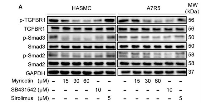
Application: WB Species: Sample: VSMCs

Application: WB Species: Human Sample: VSMCs
Application: WB Species: Mouse Sample: CFs
Application: WB Species: Mouse Sample:
Application: WB Species: Mouse Sample: Mlg cells
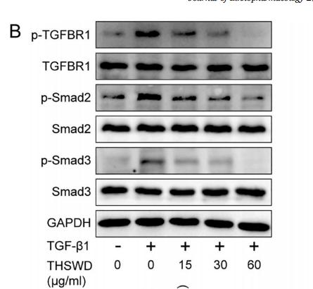
Application: WB Species: mice Sample: cardiac fibroblasts
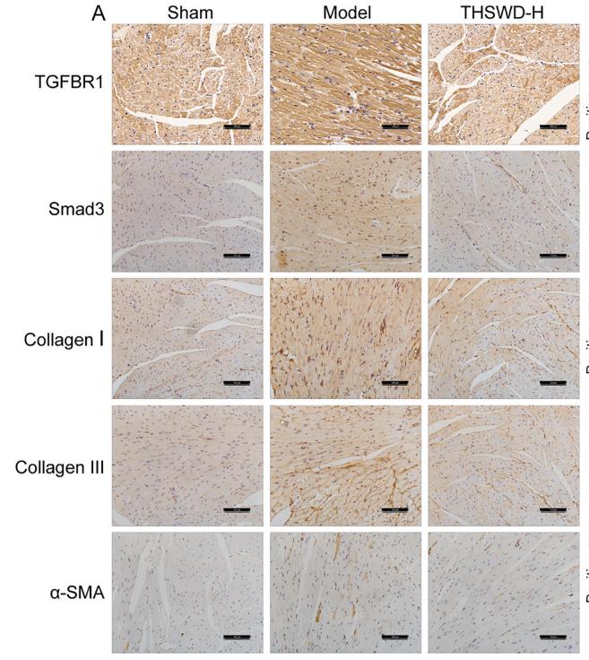
Application: IHC Species: mice Sample: heart tissues

Application: WB Species: Mice Sample: kidneys
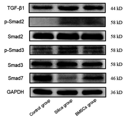
Application: WB Species: rat Sample: lung
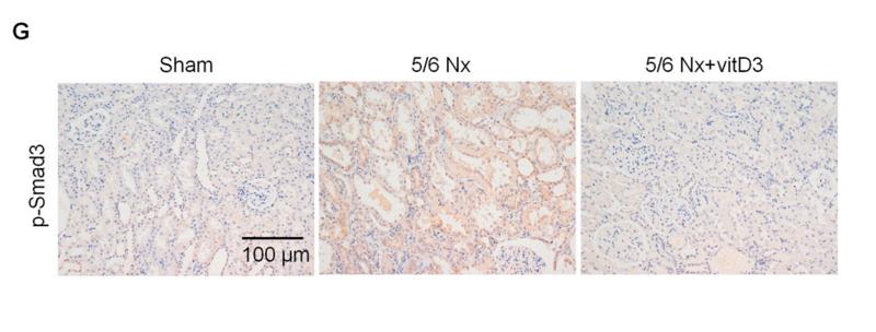
Application: IHC Species: Rat Sample: kidney tissues
Application: WB Species: human Sample: HK-2 cells
Restrictive clause
Affinity Biosciences tests all products strictly. Citations are provided as a resource for additional applications that have not been validated by Affinity Biosciences. Please choose the appropriate format for each application and consult Materials and Methods sections for additional details about the use of any product in these publications.
For Research Use Only.
Not for use in diagnostic or therapeutic procedures. Not for resale. Not for distribution without written consent. Affinity Biosciences will not be held responsible for patent infringement or other violations that may occur with the use of our products. Affinity Biosciences, Affinity Biosciences Logo and all other trademarks are the property of Affinity Biosciences LTD.