VEGFR1 Antibody - #AF6204

Product Info
*The optimal dilutions should be determined by the end user.
*Tips:
WB: For western blot detection of denatured protein samples. IHC: For immunohistochemical detection of paraffin sections (IHC-p) or frozen sections (IHC-f) of tissue samples. IF/ICC: For immunofluorescence detection of cell samples. ELISA(peptide): For ELISA detection of antigenic peptide.
Cite Format: Affinity Biosciences Cat# AF6204, RRID:AB_2835085.
Fold/Unfold
EC 2.7.10.1; FLT 1; FLT; Flt-1; FLT1; Fms like tyrosine kinase 1; Fms related tyrosine kinase 1; Fms related tyrosine kinase 1 (vascular endothelial growth factor/vascular permeability factor receptor); Fms related tyrosine kinase 1 vascular endothelial growth factor/vascular permeability factor receptor; Fms-like tyrosine kinase 1; FRT; Soluble VEGF receptor 1 14; Soluble VEGFR1 variant 2; Soluble VEGFR1 variant 21; Tyrosine protein kinase FRT; Tyrosine protein kinase receptor FLT; Tyrosine-protein kinase FRT; Tyrosine-protein kinase receptor FLT; Vascular endothelial growth factor receptor 1; Vascular endothelial growth factor vascular permeability factor receptor; Vascular permeability factor receptor 1; Vascular permeability factor receptor; VEGFR 1; VEGFR-1; VEGFR1; VGFR1_HUMAN;
Immunogens
Detected in normal lung, but also in placenta, liver, kidney, heart and brain tissues. Specifically expressed in most of the vascular endothelial cells, and also expressed in peripheral blood monocytes. Isoform 2 is strongly expressed in placenta. Isoform 3 is expressed in corneal epithelial cells (at protein level). Isoform 3 is expressed in vascular smooth muscle cells (VSMC).
- P17948 VGFR1_HUMAN:
- Protein BLAST With
- NCBI/
- ExPASy/
- Uniprot
MVSYWDTGVLLCALLSCLLLTGSSSGSKLKDPELSLKGTQHIMQAGQTLHLQCRGEAAHKWSLPEMVSKESERLSITKSACGRNGKQFCSTLTLNTAQANHTGFYSCKYLAVPTSKKKETESAIYIFISDTGRPFVEMYSEIPEIIHMTEGRELVIPCRVTSPNITVTLKKFPLDTLIPDGKRIIWDSRKGFIISNATYKEIGLLTCEATVNGHLYKTNYLTHRQTNTIIDVQISTPRPVKLLRGHTLVLNCTATTPLNTRVQMTWSYPDEKNKRASVRRRIDQSNSHANIFYSVLTIDKMQNKDKGLYTCRVRSGPSFKSVNTSVHIYDKAFITVKHRKQQVLETVAGKRSYRLSMKVKAFPSPEVVWLKDGLPATEKSARYLTRGYSLIIKDVTEEDAGNYTILLSIKQSNVFKNLTATLIVNVKPQIYEKAVSSFPDPALYPLGSRQILTCTAYGIPQPTIKWFWHPCNHNHSEARCDFCSNNEESFILDADSNMGNRIESITQRMAIIEGKNKMASTLVVADSRISGIYICIASNKVGTVGRNISFYITDVPNGFHVNLEKMPTEGEDLKLSCTVNKFLYRDVTWILLRTVNNRTMHYSISKQKMAITKEHSITLNLTIMNVSLQDSGTYACRARNVYTGEEILQKKEITIRDQEAPYLLRNLSDHTVAISSSTTLDCHANGVPEPQITWFKNNHKIQQEPGIILGPGSSTLFIERVTEEDEGVYHCKATNQKGSVESSAYLTVQGTSDKSNLELITLTCTCVAATLFWLLLTLFIRKMKRSSSEIKTDYLSIIMDPDEVPLDEQCERLPYDASKWEFARERLKLGKSLGRGAFGKVVQASAFGIKKSPTCRTVAVKMLKEGATASEYKALMTELKILTHIGHHLNVVNLLGACTKQGGPLMVIVEYCKYGNLSNYLKSKRDLFFLNKDAALHMEPKKEKMEPGLEQGKKPRLDSVTSSESFASSGFQEDKSLSDVEEEEDSDGFYKEPITMEDLISYSFQVARGMEFLSSRKCIHRDLAARNILLSENNVVKICDFGLARDIYKNPDYVRKGDTRLPLKWMAPESIFDKIYSTKSDVWSYGVLLWEIFSLGGSPYPGVQMDEDFCSRLREGMRMRAPEYSTPEIYQIMLDCWHRDPKERPRFAELVEKLGDLLQANVQQDGKDYIPINAILTGNSGFTYSTPAFSEDFFKESISAPKFNSGSSDDVRYVNAFKFMSLERIKTFEELLPNATSMFDDYQGDSSTLLASPMLKRFTWTDSKPKASLKIDLRVTSKSKESGLSDVSRPSFCHSSCGHVSEGKRRFTYDHAELERKIACCSPPPDYNSVVLYSTPPI
Predictions
Score>80(red) has high confidence and is suggested to be used for WB detection. *The prediction model is mainly based on the alignment of immunogen sequences, the results are for reference only, not as the basis of quality assurance.
High(score>80) Medium(80>score>50) Low(score<50) No confidence
PTMs - P17948 As Substrate
| Site | PTM Type | Enzyme | Source |
|---|---|---|---|
| T218 | Phosphorylation | Uniprot | |
| T222 | Phosphorylation | Uniprot | |
| T255 | Phosphorylation | Uniprot | |
| T265 | Phosphorylation | Uniprot | |
| S267 | Phosphorylation | Uniprot | |
| K306 | Methylation | Uniprot | |
| Y383 | Phosphorylation | Uniprot | |
| Y388 | Phosphorylation | Uniprot | |
| T404 | Phosphorylation | Uniprot | |
| K410 | Ubiquitination | Uniprot | |
| T588 | Phosphorylation | Uniprot | |
| T599 | Phosphorylation | Uniprot | |
| T618 | Phosphorylation | Uniprot | |
| S631 | Phosphorylation | Uniprot | |
| T633 | Phosphorylation | Uniprot | |
| S739 | Phosphorylation | Uniprot | |
| S742 | Phosphorylation | Uniprot | |
| S743 | Phosphorylation | Uniprot | |
| Y745 | Phosphorylation | Uniprot | |
| T747 | Phosphorylation | Uniprot | |
| T751 | Phosphorylation | Uniprot | |
| Y794 | Phosphorylation | P17948 (FLT1) | Uniprot |
| Y815 | Phosphorylation | Uniprot | |
| K831 | Methylation | Uniprot | |
| Y911 | Phosphorylation | Uniprot | |
| Y914 | Phosphorylation | P17948 (FLT1) | Uniprot |
| Y920 | Phosphorylation | Uniprot | |
| K932 | Methylation | Uniprot | |
| Y1048 | Phosphorylation | Uniprot | |
| Y1053 | Phosphorylation | Uniprot | |
| K1064 | Ubiquitination | Uniprot | |
| K1153 | Methylation | Uniprot | |
| Y1169 | Phosphorylation | P17948 (FLT1) | Uniprot |
| S1197 | Phosphorylation | Uniprot | |
| S1199 | Phosphorylation | Uniprot | |
| K1202 | Ubiquitination | Uniprot | |
| S1205 | Phosphorylation | Uniprot | |
| S1207 | Phosphorylation | Uniprot | |
| Y1213 | Phosphorylation | P17948 (FLT1) | Uniprot |
| Y1242 | Phosphorylation | P17948 (FLT1) | Uniprot |
| Y1309 | Phosphorylation | P17948 (FLT1) | Uniprot |
| Y1327 | Phosphorylation | P17948 (FLT1) | Uniprot |
| Y1333 | Phosphorylation | P17948 (FLT1) | Uniprot |
PTMs - P17948 As Enzyme
Research Backgrounds
Tyrosine-protein kinase that acts as a cell-surface receptor for VEGFA, VEGFB and PGF, and plays an essential role in the development of embryonic vasculature, the regulation of angiogenesis, cell survival, cell migration, macrophage function, chemotaxis, and cancer cell invasion. May play an essential role as a negative regulator of embryonic angiogenesis by inhibiting excessive proliferation of endothelial cells. Can promote endothelial cell proliferation, survival and angiogenesis in adulthood. Its function in promoting cell proliferation seems to be cell-type specific. Promotes PGF-mediated proliferation of endothelial cells, proliferation of some types of cancer cells, but does not promote proliferation of normal fibroblasts (in vitro). Has very high affinity for VEGFA and relatively low protein kinase activity; may function as a negative regulator of VEGFA signaling by limiting the amount of free VEGFA and preventing its binding to KDR. Likewise, isoforms lacking a transmembrane domain, such as isoform 2, isoform 3 and isoform 4, may function as decoy receptors for VEGFA. Modulates KDR signaling by forming heterodimers with KDR. Ligand binding leads to the activation of several signaling cascades. Activation of PLCG leads to the production of the cellular signaling molecules diacylglycerol and inositol 1,4,5-trisphosphate and the activation of protein kinase C. Mediates phosphorylation of PIK3R1, the regulatory subunit of phosphatidylinositol 3-kinase, leading to activation of phosphatidylinositol kinase and the downstream signaling pathway. Mediates activation of MAPK1/ERK2, MAPK3/ERK1 and the MAP kinase signaling pathway, as well as of the AKT1 signaling pathway. Phosphorylates SRC and YES1, and may also phosphorylate CBL. Isoform 1 phosphorylates PLCG. Promotes phosphorylation of AKT1 at 'Ser-473'. Promotes phosphorylation of PTK2/FAK1. Isoform 7 has a truncated kinase domain; it increases phosphorylation of SRC at 'Tyr-418' by unknown means and promotes tumor cell invasion.
N-glycosylated.
Ubiquitinated after VEGFA-mediated autophosphorylation, leading to proteolytic degradation.
Autophosphorylated on tyrosine residues upon ligand binding. Autophosphorylation occurs in trans, i.e. one subunit of the dimeric receptor phosphorylates tyrosine residues on the other subunit. Phosphorylation at Tyr-1169 is important for interaction with PLCG. Phosphorylation at Tyr-1213 is important for interaction with PIK3R1, PTPN11, GRB2, and PLCG. Phosphorylation at Tyr-1333 is important for endocytosis and for interaction with CBL, NCK1 and CRK. Is probably dephosphorylated by PTPRB.
Cell membrane>Single-pass type I membrane protein. Endosome.
Note: Autophosphorylation promotes ubiquitination and endocytosis.
Secreted.
Secreted.
Secreted.
Cytoplasm.
Cytoplasm.
Cytoplasm.
Detected in normal lung, but also in placenta, liver, kidney, heart and brain tissues. Specifically expressed in most of the vascular endothelial cells, and also expressed in peripheral blood monocytes. Isoform 2 is strongly expressed in placenta. Isoform 3 is expressed in corneal epithelial cells (at protein level). Isoform 3 is expressed in vascular smooth muscle cells (VSMC).
Interacts with VEGFA, VEGFB and PGF. Monomer in the absence of bound VEGFA, VEGFB or PGF. Homodimer in the presence of bound VEGFA, VEGFB and PGF. Can also form a heterodimer with KDR. Interacts (when tyrosine phosphorylated) with CBL, CRK, GRB2, NCK1, PIK3R1, PLCG, PSEN1 and PTPN11. Probably interacts also with PTPRB. Interacts with RACK1. Identified in a complex with CBL and CD2AP.
The second and third Ig-like C2-type (immunoglobulin-like) domains are sufficient for VEGFA binding.
Belongs to the protein kinase superfamily. Tyr protein kinase family. CSF-1/PDGF receptor subfamily.
Research Fields
· Cellular Processes > Cellular community - eukaryotes > Focal adhesion. (View pathway)
· Environmental Information Processing > Signal transduction > MAPK signaling pathway. (View pathway)
· Environmental Information Processing > Signal transduction > Ras signaling pathway. (View pathway)
· Environmental Information Processing > Signal transduction > Rap1 signaling pathway. (View pathway)
· Environmental Information Processing > Signaling molecules and interaction > Cytokine-cytokine receptor interaction. (View pathway)
· Environmental Information Processing > Signal transduction > HIF-1 signaling pathway. (View pathway)
· Environmental Information Processing > Signal transduction > PI3K-Akt signaling pathway. (View pathway)
· Human Diseases > Cancers: Overview > Transcriptional misregulation in cancer.
· Human Diseases > Immune diseases > Rheumatoid arthritis.
References
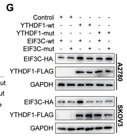
Application: WB Species: human Sample: PLC-PRF-5 cells

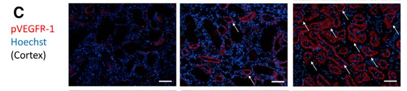
Application: IHC Species: human Sample: HCC
Application: WB Species: human Sample: PLC-PRF-5 cells
Application: IF/ICC Species: rat Sample: Bone marrow cells
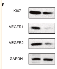
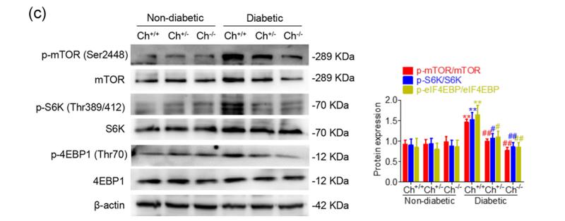
Application: WB Species: Mouse Sample:
Application: WB Species: human Sample: bone marrow
Restrictive clause
Affinity Biosciences tests all products strictly. Citations are provided as a resource for additional applications that have not been validated by Affinity Biosciences. Please choose the appropriate format for each application and consult Materials and Methods sections for additional details about the use of any product in these publications.
For Research Use Only.
Not for use in diagnostic or therapeutic procedures. Not for resale. Not for distribution without written consent. Affinity Biosciences will not be held responsible for patent infringement or other violations that may occur with the use of our products. Affinity Biosciences, Affinity Biosciences Logo and all other trademarks are the property of Affinity Biosciences LTD.