p57 Kip2 Antibody - #AF6819
, blocked with antigen-specific peptides,
Lane 2: Hela cells(serum starvation treatment),
Lane 3: HepG2 cells(heat shock treatment).")
| Product: | p57 Kip2 Antibody |
| Catalog: | AF6819 |
| Description: | Rabbit polyclonal antibody to p57 Kip2 |
| Application: | WB |
| Reactivity: | Human |
| Mol.Wt.: | 32kD(Calculated). |
| Uniprot: | P49918 |
| RRID: | AB_2847542 |
Product Info
*The optimal dilutions should be determined by the end user.
*Tips:
WB: For western blot detection of denatured protein samples. IHC: For immunohistochemical detection of paraffin sections (IHC-p) or frozen sections (IHC-f) of tissue samples. IF/ICC: For immunofluorescence detection of cell samples. ELISA(peptide): For ELISA detection of antigenic peptide.
Cite Format: Affinity Biosciences Cat# AF6819, RRID:AB_2847542.
Fold/Unfold
Beckwith Wiedemann syndrome; BWCR; BWS; CDKI; CDKN 1C; CDKN1C; CDN1C_HUMAN; Cyclin dependent kinase inhibitor 1C; Cyclin dependent kinase inhibitor p57; Cyclin-dependent kinase inhibitor 1C; Cyclin-dependent kinase inhibitor p57; KIP 2; KIP2; p57; p57 Kip 2; p57KIP2; WBS;
Immunogens
A synthesized peptide derived from human p57 Kip2.
Expressed in the heart, brain, lung, skeletal muscle, kidney, pancreas and testis. Expressed in the eye. High levels are seen in the placenta while low levels are seen in the liver.
- P49918 CDN1C_HUMAN:
- Protein BLAST With
- NCBI/
- ExPASy/
- Uniprot
MSDASLRSTSTMERLVARGTFPVLVRTSACRSLFGPVDHEELSRELQARLAELNAEDQNRWDYDFQQDMPLRGPGRLQWTEVDSDSVPAFYRETVQVGRCRLLLAPRPVAVAVAVSPPLEPAAESLDGLEEAPEQLPSVPVPAPASTPPPVPVLAPAPAPAPAPVAAPVAAPVAVAVLAPAPAPAPAPAPAPAPVAAPAPAPAPAPAPAPAPAPAPDAAPQESAEQGANQGQRGQEPLADQLHSGISGRPAAGTAAASANGAAIKKLSGPLISDFFAKRKRSAPEKSSGDVPAPCPSPSAAPGVGSVEQTPRKRLR
PTMs - P49918 As Substrate
| Site | PTM Type | Enzyme | Source |
|---|---|---|---|
| S43 | Phosphorylation | Uniprot | |
| S146 | Phosphorylation | Q16539 (MAPK14) | Uniprot |
| K266 | Acetylation | Uniprot | |
| S268 | Phosphorylation | Uniprot | |
| K278 | Acetylation | Uniprot | |
| S282 | Phosphorylation | P31749 (AKT1) | Uniprot |
| S297 | Phosphorylation | Uniprot | |
| S299 | Phosphorylation | Uniprot | |
| S306 | Phosphorylation | Uniprot | |
| T310 | Phosphorylation | P31749 (AKT1) | Uniprot |
Research Backgrounds
Potent tight-binding inhibitor of several G1 cyclin/CDK complexes (cyclin E-CDK2, cyclin D2-CDK4, and cyclin A-CDK2) and, to lesser extent, of the mitotic cyclin B-CDC2. Negative regulator of cell proliferation. May play a role in maintenance of the non-proliferative state throughout life.
Nucleus.
Expressed in the heart, brain, lung, skeletal muscle, kidney, pancreas and testis. Expressed in the eye. High levels are seen in the placenta while low levels are seen in the liver.
Interacts with PCNA.
Belongs to the CDI family.
Research Fields
· Cellular Processes > Cell growth and death > Cell cycle. (View pathway)
References
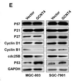
Application: WB Species: Human Sample: gastric cancer cell
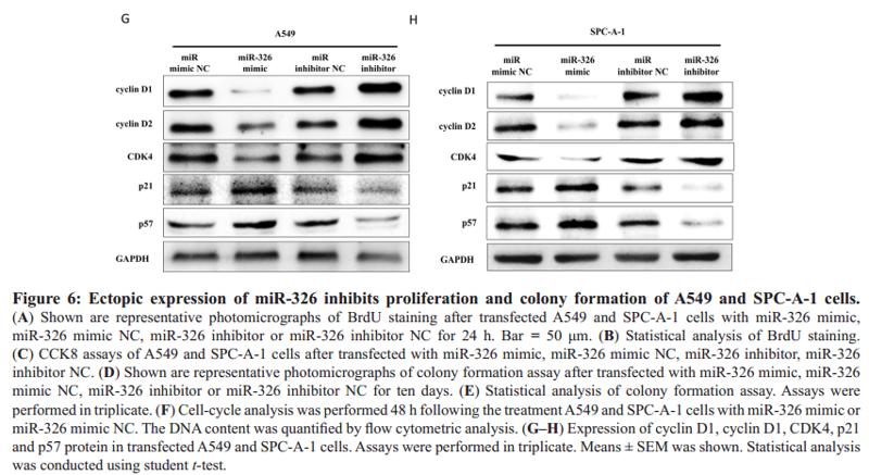
Application: WB Species: human Sample: A549 cells
Application: WB Species: Sample:
Restrictive clause
Affinity Biosciences tests all products strictly. Citations are provided as a resource for additional applications that have not been validated by Affinity Biosciences. Please choose the appropriate format for each application and consult Materials and Methods sections for additional details about the use of any product in these publications.
For Research Use Only.
Not for use in diagnostic or therapeutic procedures. Not for resale. Not for distribution without written consent. Affinity Biosciences will not be held responsible for patent infringement or other violations that may occur with the use of our products. Affinity Biosciences, Affinity Biosciences Logo and all other trademarks are the property of Affinity Biosciences LTD.