SRF Antibody - #AF6160
 and mouse anti-beta tubulin Ab(#T0023) for 1 hour at 37°C. An AlexaFluor594 conjugated goat anti-rabbit IgG Ab(Red) and an AlexaFluor488 conjugated goat anti-mouse IgG Ab(Green) were used as the secondary antibody.
The nuclear counter stain is DAPI (blue).")
| Product: | SRF Antibody |
| Catalog: | AF6160 |
| Description: | Rabbit polyclonal antibody to SRF |
| Application: | WB IHC IF/ICC |
| Reactivity: | Human, Mouse, Rat |
| Prediction: | Pig, Bovine, Rabbit, Dog |
| Mol.Wt.: | 67kDa; 52kD(Calculated). |
| Uniprot: | P11831 |
| RRID: | AB_2835029 |
Product Info
*The optimal dilutions should be determined by the end user.
*Tips:
WB: For western blot detection of denatured protein samples. IHC: For immunohistochemical detection of paraffin sections (IHC-p) or frozen sections (IHC-f) of tissue samples. IF/ICC: For immunofluorescence detection of cell samples. ELISA(peptide): For ELISA detection of antigenic peptide.
Cite Format: Affinity Biosciences Cat# AF6160, RRID:AB_2835029.
Fold/Unfold
c fos serum response element binding factor; c fos serum response element binding transcription factor; ELK3; ERP; MCM 1; MCM1; OTTHUMP00000039820; SAP2; Serum response factor; SRF; SRF serum response factor c fos serum response element binding transcription factor; SRF_HUMAN;
Immunogens
- P11831 SRF_HUMAN:
- Protein BLAST With
- NCBI/
- ExPASy/
- Uniprot
MLPTQAGAAAALGRGSALGGSLNRTPTGRPGGGGGTRGANGGRVPGNGAGLGPGRLEREAAAAAATTPAPTAGALYSGSEGDSESGEEEELGAERRGLKRSLSEMEIGMVVGGPEASAAATGGYGPVSGAVSGAKPGKKTRGRVKIKMEFIDNKLRRYTTFSKRKTGIMKKAYELSTLTGTQVLLLVASETGHVYTFATRKLQPMITSETGKALIQTCLNSPDSPPRSDPTTDQRMSATGFEETDLTYQVSESDSSGETKDTLKPAFTVTNLPGTTSTIQTAPSTSTTMQVSSGPSFPITNYLAPVSASVSPSAVSSANGTVLKSTGSGPVSSGGLMQLPTSFTLMPGGAVAQQVPVQAIQVHQAPQQASPSRDSSTDLTQTSSSGTVTLPATIMTSSVPTTVGGHMMYPSPHAVMYAPTSGLGDGSLTVLNAFSQAPSTMQVSHSQVQEPGGVPQVFLTASSGTVQIPVSAVQLHQMAVIGQQAGSSSNLTELQVVNLDTAHSTKSE
Predictions
Score>80(red) has high confidence and is suggested to be used for WB detection. *The prediction model is mainly based on the alignment of immunogen sequences, the results are for reference only, not as the basis of quality assurance.
High(score>80) Medium(80>score>50) Low(score<50) No confidence
PTMs - P11831 As Substrate
| Site | PTM Type | Enzyme | Source |
|---|---|---|---|
| T4 | Phosphorylation | Uniprot | |
| R14 | Methylation | Uniprot | |
| S16 | Phosphorylation | Uniprot | |
| S21 | Phosphorylation | Uniprot | |
| R24 | Methylation | Uniprot | |
| T27 | Phosphorylation | Uniprot | |
| R37 | Methylation | Uniprot | |
| T71 | Phosphorylation | Uniprot | |
| Y76 | Phosphorylation | Uniprot | |
| S77 | Phosphorylation | P68400 (CSNK2A1) | Uniprot |
| S79 | Phosphorylation | P68400 (CSNK2A1) | Uniprot |
| S83 | Phosphorylation | P68400 (CSNK2A1) | Uniprot |
| S85 | Phosphorylation | P49137 (MAPKAPK2) , P68400 (CSNK2A1) | Uniprot |
| S101 | Phosphorylation | Uniprot | |
| S103 | Phosphorylation | Q15418 (RPS6KA1) , O75582 (RPS6KA5) , Q9UQM7 (CAMK2A) , P49137 (MAPKAPK2) | Uniprot |
| K147 | Sumoylation | Uniprot | |
| T159 | Phosphorylation | P17612 (PRKACA) , Q13976 (PRKG1) , Q09013 (DMPK) , P17252 (PRKCA) | Uniprot |
| T160 | Phosphorylation | Q9UQM7 (CAMK2A) , Q05655 (PRKCD) | Uniprot |
| S162 | Phosphorylation | P17252 (PRKCA) | Uniprot |
| S176 | Phosphorylation | Uniprot | |
| T177 | Phosphorylation | Uniprot | |
| T199 | Phosphorylation | Uniprot | |
| S221 | Phosphorylation | Uniprot | |
| S224 | Phosphorylation | Uniprot | |
| S228 | Phosphorylation | Uniprot | |
| S251 | Phosphorylation | Uniprot | |
| S253 | Phosphorylation | Uniprot | |
| S277 | O-Glycosylation | Uniprot | |
| S307 | O-Glycosylation | Uniprot | |
| S309 | O-Glycosylation | Uniprot | |
| S313 | O-Glycosylation | Uniprot | |
| S316 | O-Glycosylation | Uniprot | |
| S370 | Phosphorylation | Uniprot | |
| S383 | O-Glycosylation | Uniprot | |
| T401 | O-Glycosylation | Uniprot | |
| S435 | Phosphorylation | P78527 (PRKDC) | Uniprot |
| S446 | Phosphorylation | P78527 (PRKDC) | Uniprot |
Research Backgrounds
SRF is a transcription factor that binds to the serum response element (SRE), a short sequence of dyad symmetry located 300 bp to the 5' of the site of transcription initiation of some genes (such as FOS). Together with MRTFA transcription coactivator, controls expression of genes regulating the cytoskeleton during development, morphogenesis and cell migration. The SRF-MRTFA complex activity responds to Rho GTPase-induced changes in cellular globular actin (G-actin) concentration, thereby coupling cytoskeletal gene expression to cytoskeletal dynamics. Required for cardiac differentiation and maturation.
Phosphorylated by PRKDC.
Nucleus.
Binds DNA as a multimer, probably a dimer. Interacts with MRTFA, forming the SRF-MRTFA nuclear complex which binds the 5'-CArG-3' consensus motif (CArG box) on DNA via SRF. Forms a nuclear ternary complex with MRTFA and SCAI. Interacts with MRTFB. Interacts with MLLT7/FOXO4, NKX3A and SSRP1. Interacts with ARID2 (By similarity). Interacts with SRFBP1 (By similarity). Interacts with FOXK1. Interacts with LPXN. Interacts with OLFM2; the interaction promotes dissociation of SRF from the transcriptional repressor HEY2, facilitates binding of SRF to target genes and promotes smooth muscle differentiation.
Research Fields
· Environmental Information Processing > Signal transduction > MAPK signaling pathway. (View pathway)
· Environmental Information Processing > Signal transduction > cGMP-PKG signaling pathway. (View pathway)
· Human Diseases > Infectious diseases: Viral > HTLV-I infection.
· Human Diseases > Cancers: Overview > Viral carcinogenesis.
References

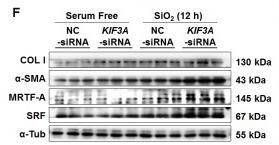
Application: WB Species: human Sample: MRC-5 fibroblasts
Application: WB Species: Human Sample: MRC-5 fibroblasts
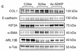
Application: WB Species: Rat Sample:
Restrictive clause
Affinity Biosciences tests all products strictly. Citations are provided as a resource for additional applications that have not been validated by Affinity Biosciences. Please choose the appropriate format for each application and consult Materials and Methods sections for additional details about the use of any product in these publications.
For Research Use Only.
Not for use in diagnostic or therapeutic procedures. Not for resale. Not for distribution without written consent. Affinity Biosciences will not be held responsible for patent infringement or other violations that may occur with the use of our products. Affinity Biosciences, Affinity Biosciences Logo and all other trademarks are the property of Affinity Biosciences LTD.