Product: Tyrosine Hydroxylase Antibody
Catalog: AF6113
Description: Rabbit polyclonal antibody to Tyrosine Hydroxylase
Application: WB IHC IF/ICC
Reactivity: Human, Mouse, Rat
Prediction: Dog
Mol.Wt.: 60kDa; 59kD(Calculated).
Uniprot: P07101
RRID: AB_2834999

View similar products>>

   Size Price Inventory
 100ul $280 In stock
 200ul $350 In stock

Lead Time: Same day delivery

For pricing and ordering contact:
Local distributors

Product Info

Source:
Rabbit
Application:
WB 1:500-1:2000, IHC 1:50-1:200, IF/ICC 1:100-1:500
*The optimal dilutions should be determined by the end user.
*Tips:

WB: For western blot detection of denatured protein samples. IHC: For immunohistochemical detection of paraffin sections (IHC-p) or frozen sections (IHC-f) of tissue samples. IF/ICC: For immunofluorescence detection of cell samples. ELISA(peptide): For ELISA detection of antigenic peptide.

Reactivity:
Human,Mouse,Rat
Prediction:
Dog(82%)
Clonality:
Polyclonal
Specificity:
Tyrosine Hydroxylase Antibody detects endogenous levels of total Tyrosine Hydroxylase.
RRID:
AB_2834999
Cite Format: Affinity Biosciences Cat# AF6113, RRID:AB_2834999.
Conjugate:
Unconjugated.
Purification:
The antiserum was purified by peptide affinity chromatography using SulfoLink™ Coupling Resin (Thermo Fisher Scientific).
Storage:
Rabbit IgG in phosphate buffered saline , pH 7.4, 150mM NaCl, 0.02% sodium azide and 50% glycerol. Store at -20 °C. Stable for 12 months from date of receipt.
Alias:

Fold/Unfold

Dystonia 14; DYT14; DYT5b; EC 1.14.16.2; OTTHUMP00000011225; OTTHUMP00000011226; ple; Protein Pale; TH; The; TY3H_HUMAN; TYH; Tyrosine 3 hydroxylase; Tyrosine 3 monooxygenase; Tyrosine 3-hydroxylase; Tyrosine 3-monooxygenase; Tyrosine hydroxylase;

Immunogens

Immunogen:
Uniprot:
Gene(ID):
Expression:
P07101 TY3H_HUMAN:

Mainly expressed in the brain and adrenal glands.

Description:
Tyrosine hydroxylase (EC 1.14.16.2) is involved in the conversion of phenylalanine to dopamine. As the rate-limiting enzyme in the synthesis of catecholamines, tyrosine hydroxylase has a key role in the physiology of adrenergic neurons.
Sequence:
MPTPDATTPQAKGFRRAVSELDAKQAEAIMVRGQGAPGPSLTGSPWPGTAAPAASYTPTPRSPRFIGRRQSLIEDARKEREAAVAAAAAAVPSEPGDPLEAVAFEEKEGKAVLNLLFSPRATKPSALSRAVKVFETFEAKIHHLETRPAQRPRAGGPHLEYFVRLEVRRGDLAALLSGVRQVSEDVRSPAGPKVPWFPRKVSELDKCHHLVTKFDPDLDLDHPGFSDQVYRQRRKLIAEIAFQYRHGDPIPRVEYTAEEIATWKEVYTTLKGLYATHACGEHLEAFALLERFSGYREDNIPQLEDVSRFLKERTGFQLRPVAGLLSARDFLASLAFRVFQCTQYIRHASSPMHSPEPDCCHELLGHVPMLADRTFAQFSQDIGLASLGASDEEIEKLSTLYWFTVEFGLCKQNGEVKAYGAGLLSSYGELLHCLSEEPEIRAFDPEAAAVQPYQDQTYQSVYFVSESFSDAKDKLRSYASRIQRPFSVKFDPYTLAIDVLDSPQAVRRSLEGVQDELDTLAHALSAIG

Predictions

Predictions:

Score>80(red) has high confidence and is suggested to be used for WB detection. *The prediction model is mainly based on the alignment of immunogen sequences, the results are for reference only, not as the basis of quality assurance.

Species
Results
Score
Dog
82
Bovine
73
Xenopus
73
Rabbit
73
Zebrafish
27
Pig
0
Horse
0
Sheep
0
Chicken
0
Model Confidence:
High(score>80) Medium(80>score>50) Low(score<50) No confidence

PTMs - P07101 As Substrate

Site PTM Type Enzyme
T3 Phosphorylation
S19 Phosphorylation Q8IW41 (MAPKAPK5) , P49137 (MAPKAPK2) , Q9UQM7 (CAMK2A)
S40 Phosphorylation P49137 (MAPKAPK2) , P51812 (RPS6KA3) , O75582 (RPS6KA5)
S62 Phosphorylation P28482 (MAPK1) , P27361 (MAPK3)
S71 Phosphorylation P49137 (MAPKAPK2) , P51812 (RPS6KA3) , P17612 (PRKACA) , Q9UQM7 (CAMK2A)
S118 Phosphorylation
Y230 Phosphorylation
Y419 Phosphorylation
S502 Phosphorylation

Research Backgrounds

Function:

Plays an important role in the physiology of adrenergic neurons.

Tissue Specificity:

Mainly expressed in the brain and adrenal glands.

Family&Domains:

Belongs to the biopterin-dependent aromatic amino acid hydroxylase family.

Research Fields

· Human Diseases > Neurodegenerative diseases > Parkinson's disease.

· Human Diseases > Substance dependence > Cocaine addiction.

· Human Diseases > Substance dependence > Amphetamine addiction.

· Human Diseases > Substance dependence > Alcoholism.

· Metabolism > Amino acid metabolism > Tyrosine metabolism.

· Metabolism > Metabolism of cofactors and vitamins > Folate biosynthesis.

· Metabolism > Global and overview maps > Metabolic pathways.

· Organismal Systems > Nervous system > Dopaminergic synapse.

· Organismal Systems > Endocrine system > Prolactin signaling pathway.   (View pathway)

References

1). Lipopolysaccharide-binding protein expression is increased by stress and inhibits monoamine synthesis to promote depressive symptoms. Immunity (PubMed: 36854305) [IF=32.4]

2). Exposure to deltamethrin in adolescent mice induced thyroid dysfunction and behavioral disorders. Chemosphere (PubMed: 31683416) [IF=8.8]

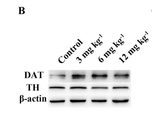
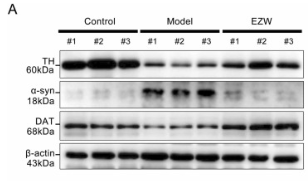
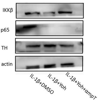
Application: WB    Species: Mice    Sample: striatum tissue

Fig. 5. DM damaged thyroid histopathology and protein expression of DAT and TH. (A) Representative images of thyroid gland stained with HE. Asterisk: expanded thyroid follicle. Solid arrowheads: hyperplastic thyroid follicular epithelial cells. Arrows: shed thyroid follicular epithelial cells. (B) Western blotting analysis the levels of DAT and TH in striatum tissue. (C) The statistical result of DAT protein analysis. (D) The statistical result of TH protein analysis. **p < 0.01, ****P < 0.0001.

3). Integrative multilevel exploration of the mechanism by which Er-Zhi-Wan alleviates the Parkinson's disease (PD)-like phenotype in the MPTP-induced PD mouse model. Biomedicine & Pharmacotherapy (PubMed: 37348406) [IF=7.5]

Application: WB    Species: Mouse    Sample:

Fig. 3. EZW reduces the dopaminergic neuron damage in MPTP-PD mice. (A) The expression of TH, α-syn, and DAT in the striatum of each group was measured by Western blot analysis (n = 3 per group). (B) Relative densities of TH, α-syn, DAT, and β-actin were measured in different groups using densitometric analysis of Western blots. TH, α-syn, and DAT proteins were normalized to β-actin, and were expressed as the fold difference relative to the control group (Data represent the mean ± SD, n = 3. * P 

4). SiNiSan ameliorates depression-like behavior in rats by enhancing synaptic plasticity via the CaSR-PKC-ERK signaling pathway. BIOMEDICINE & PHARMACOTHERAPY (PubMed: 31958763) [IF=7.5]

Application: IHC    Species: rat    Sample: hippocampus

Fig. 4. |Effects of SNS on the Syn protein in the HIP and PFC of stressed rats. (A–D). Syn immunostaining and quantification in the CA1 and PFC. (A–B): Quantitative analysis of IHC in hippocampal CA1 and PFC regions. (C–D): Representative IHC figures of CA1 and PFC, scale bar = 50 μm. All data are expressed as mean ± S.E.M.(*p < 0.05, **p < 0.01 vs. the model group).

5). Preparation of levodopa-loaded crystalsomes through thermally induced crystallization reverses functional deficits in Parkinsonian mice. Biomaterials Science (PubMed: 30702723) [IF=6.6]

Application: WB    Species: mouse    Sample: striatal

Figure 5.| Immunohistochemistry and western bolt of TH expression in striatal. a. Immunohistochemistry staining of brain exhibited the expression of TH in striatal. Black dotted box presented the area of striatum. The results showed that LD crystalsomes treatment enhanced the expression of TH in striatal. b. Western bolt for TH relative expression of striatal. Data are presented as means ± standard error (n=6). Tukey’s multiple comparison test, * p < 0.05, ** p < 0.01, *** p < 0.001.

6). Chitosan oligosaccharides exert neuroprotective effects via modulating the PI3K/Akt/Bcl-2 pathway in a Parkinsonian model. Food & Function (PubMed: 35545086) [IF=6.1]

7). Oligo-Porphyran Ameliorates Neurobehavioral Deficits in Parkinsonian Mice by Regulating the PI3K/Akt/Bcl-2 Pathway. Marine Drugs (PubMed: 29509717) [IF=5.4]

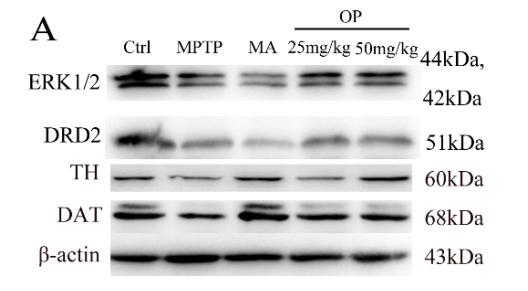
Application: WB    Species: mouse    Sample: striatum

Figure 3. |Effects of OP on the levels of expression of extracellular signal-regulated protein kinases 1 and 2 (ERK1/2), dopamine receptor D2 (DRD2), tyrosine hydroxylase (TH), and dopamine transporter(DAT) in the striatum. After pretreated with MPTP for seven days, the C57BL/6 mice were administrated with MA or different concentrations of OP for the followed seven days.(A): Original bands of ERK1/2, DRD2, TH, DAT, and β-actin.

8). Yohimbine Ameliorates Temporomandibular Joint Chondrocyte Inflammation with Suppression of NF-κB Pathway. INFLAMMATION (PubMed: 32789555) [IF=5.1]

Application: WB    Species: Mice    Sample:

Fig. 5. Reversal of the effects of Yoh by a tyrosine hydroxylase inhibitor through the NF-κB pathway. (a) Western blot showing TH expression in the normal and IL-1β groups. Immunohistochemical staining of TH in the condylar process in the (b) normal and elevated-occlusion groups and (c) sham and surgery groups. Western blot showing the expression of TH in the (d) normal, IL-1β + DMSO, and IL-1β + Yoh groups and (e) IL-1β + DMSO, IL-1β + Yoh, and IL-1β + Yoh + αmpT groups. (f) Cytometric bead array showing the expression of IL-6 in the IL-1β + DMSO, IL-1β + Yoh, and IL-1β + Yoh + αmpT groups.*P < 0.05 compared with the control group, **P < 0.01 compared with the control group. Yoh, Yohimbine; TH, tyrosine hydroxylase.

9). Acetyl‐L‐carnitine improves erectile function in bilateral cavernous nerve injury rats via promoting cavernous nerve regeneration. Andrology (PubMed: 35420721) [IF=4.5]

10). Polydatin alleviates parkinsonism in MPTP-model mice by enhancing glycolysis in dopaminergic neurons. Neurochemistry International (PubMed: 32758587) [IF=4.2]

Load more

Restrictive clause


Affinity Biosciences tests all products strictly. Citations are provided as a resource for additional applications that have not been validated by Affinity Biosciences. Please choose the appropriate format for each application and consult Materials and Methods sections for additional details about the use of any product in these publications.

For Research Use Only.
Not for use in diagnostic or therapeutic procedures. Not for resale. Not for distribution without written consent. Affinity Biosciences will not be held responsible for patent infringement or other violations that may occur with the use of our products. Affinity Biosciences, Affinity Biosciences Logo and all other trademarks are the property of Affinity Biosciences LTD.