Product: ICAM1 Antibody
Catalog: AF6088
Description: Rabbit polyclonal antibody to ICAM1
Application: WB IHC IF/ICC
Reactivity: Human, Mouse, Rat, Monkey
Prediction: Rabbit
Mol.Wt.: 92kDa, 57 kDa; 58kD(Calculated).
Uniprot: P05362
RRID: AB_2834982

View similar products>>

   Size Price Inventory
 100ul $280 In stock
 200ul $350 In stock

Lead Time: Same day delivery

For pricing and ordering contact:
Local distributors

Product Info

Source:
Rabbit
Application:
WB 1:500-1:2000, IHC 1:50-1:200, IF/ICC 1:100-1:500
*The optimal dilutions should be determined by the end user.
*Tips:

WB: For western blot detection of denatured protein samples. IHC: For immunohistochemical detection of paraffin sections (IHC-p) or frozen sections (IHC-f) of tissue samples. IF/ICC: For immunofluorescence detection of cell samples. ELISA(peptide): For ELISA detection of antigenic peptide.

Reactivity:
Human,Mouse,Rat,Monkey
Prediction:
Rabbit(82%)
Clonality:
Polyclonal
Specificity:
ICAM1 Antibody detects endogenous levels of total ICAM1.
RRID:
AB_2834982
Cite Format: Affinity Biosciences Cat# AF6088, RRID:AB_2834982.
Conjugate:
Unconjugated.
Purification:
The antiserum was purified by peptide affinity chromatography using SulfoLink™ Coupling Resin (Thermo Fisher Scientific).
Storage:
Rabbit IgG in phosphate buffered saline , pH 7.4, 150mM NaCl, 0.02% sodium azide and 50% glycerol. Store at -20 °C. Stable for 12 months from date of receipt.
Alias:

Fold/Unfold

Antigen identified by monoclonal antibody BB2; BB 2; BB2; CD 54; CD_antigen=CD54; CD54; Cell surface glycoprotein P3.58; Human rhinovirus receptor; ICAM 1; ICAM-1; ICAM1; ICAM1_HUMAN; intercellular adhesion molecule 1 (CD54), human rhinovirus receptor; Intercellular adhesion molecule 1; Major group rhinovirus receptor; MALA 2; MALA2; MyD 10; MyD10; P3.58; Surface antigen of activated B cells, BB2;

Immunogens

Immunogen:
Uniprot:
Gene(ID):
Description:
ICAM1 a type I membrane protein of the immunoglobulin superfamily. Is a ligand for the leukocyte adhesion LFA-1 protein (Integrin alpha-L/beta-2) and a Rhinovirus receptor. Typically expressed on endothelial cells and cells of the immune system.
Sequence:
MAPSSPRPALPALLVLLGALFPGPGNAQTSVSPSKVILPRGGSVLVTCSTSCDQPKLLGIETPLPKKELLLPGNNRKVYELSNVQEDSQPMCYSNCPDGQSTAKTFLTVYWTPERVELAPLPSWQPVGKNLTLRCQVEGGAPRANLTVVLLRGEKELKREPAVGEPAEVTTTVLVRRDHHGANFSCRTELDLRPQGLELFENTSAPYQLQTFVLPATPPQLVSPRVLEVDTQGTVVCSLDGLFPVSEAQVHLALGDQRLNPTVTYGNDSFSAKASVSVTAEDEGTQRLTCAVILGNQSQETLQTVTIYSFPAPNVILTKPEVSEGTEVTVKCEAHPRAKVTLNGVPAQPLGPRAQLLLKATPEDNGRSFSCSATLEVAGQLIHKNQTRELRVLYGPRLDERDCPGNWTWPENSQQTPMCQAWGNPLPELKCLKDGTFPLPIGESVTVTRDLEGTYLCRARSTQGEVTRKVTVNVLSPRYEIVIITVVAAAVIMGTAGLSTYLYNRQRKIKKYRLQQAQKGTPMKPNTQATPP

Predictions

Predictions:

Score>80(red) has high confidence and is suggested to be used for WB detection. *The prediction model is mainly based on the alignment of immunogen sequences, the results are for reference only, not as the basis of quality assurance.

Species
Results
Score
Rabbit
82
Pig
73
Bovine
73
Sheep
73
Dog
73
Horse
55
Xenopus
0
Zebrafish
0
Chicken
0
Model Confidence:
High(score>80) Medium(80>score>50) Low(score<50) No confidence

PTMs - P05362 As Substrate

Site PTM Type Enzyme
S43 Phosphorylation
T62 O-Glycosylation
T62 Phosphorylation
N130 N-Glycosylation
N145 N-Glycosylation
N183 N-Glycosylation
N202 N-Glycosylation
Y207 Phosphorylation
T211 Phosphorylation
T217 Phosphorylation
S223 Phosphorylation
N267 N-Glycosylation
S275 Phosphorylation
N296 N-Glycosylation
S323 Phosphorylation
K359 Ubiquitination
N385 N-Glycosylation
Y501 Phosphorylation
Y512 Phosphorylation
K519 Ubiquitination
T521 Phosphorylation
K524 Ubiquitination
T527 Phosphorylation
T530 Phosphorylation

Research Backgrounds

Function:

ICAM proteins are ligands for the leukocyte adhesion protein LFA-1 (integrin alpha-L/beta-2). During leukocyte trans-endothelial migration, ICAM1 engagement promotes the assembly of endothelial apical cups through ARHGEF26/SGEF and RHOG activation.

(Microbial infection) Acts as a receptor for major receptor group rhinovirus A-B capsid proteins.

(Microbial infection) Acts as a receptor for Coxsackievirus A21 capsid proteins.

(Microbial infection) Upon Kaposi's sarcoma-associated herpesvirus/HHV-8 infection, is degraded by viral E3 ubiquitin ligase MIR2, presumably to prevent lysis of infected cells by cytotoxic T-lymphocytes and NK cell.

PTMs:

Monoubiquitinated, which is promoted by MARCH9 and leads to endocytosis.

Subcellular Location:

Membrane>Single-pass type I membrane protein.

Extracellular region or secreted Cytosol Plasma membrane Cytoskeleton Lysosome Endosome Peroxisome ER Golgi apparatus Nucleus Mitochondrion Manual annotation Automatic computational assertionSubcellular location
Subunit Structure:

Homodimer (Probable). Interacts with MUC1 and promotes cell aggregation in epithelial cells. Interacts with ARHGEF26/SGEF. Interacts (on T cell side) with CD81, CD247 and CD9 at immunological synapses between antigen-presenting cells and T cells.

(Microbial infection) Interacts with major receptor group rhinovirus A-B capsid proteins.

(Microbial infection) Interacts with Coxsackievirus A21 capsid proteins.

Family&Domains:

Belongs to the immunoglobulin superfamily. ICAM family.

Research Fields

· Environmental Information Processing > Signal transduction > NF-kappa B signaling pathway.   (View pathway)

· Environmental Information Processing > Signaling molecules and interaction > Cell adhesion molecules (CAMs).   (View pathway)

· Environmental Information Processing > Signal transduction > TNF signaling pathway.   (View pathway)

· Human Diseases > Infectious diseases: Parasitic > African trypanosomiasis.

· Human Diseases > Infectious diseases: Parasitic > Malaria.

· Human Diseases > Infectious diseases: Bacterial > Staphylococcus aureus infection.

· Human Diseases > Infectious diseases: Viral > Influenza A.

· Human Diseases > Infectious diseases: Viral > HTLV-I infection.

· Human Diseases > Infectious diseases: Viral > Epstein-Barr virus infection.

· Human Diseases > Immune diseases > Rheumatoid arthritis.

· Human Diseases > Cardiovascular diseases > Viral myocarditis.

· Organismal Systems > Immune system > Natural killer cell mediated cytotoxicity.   (View pathway)

· Organismal Systems > Immune system > Leukocyte transendothelial migration.   (View pathway)

References

1). A novel UV-curable extravascular stent to prevent restenosis of venous grafts. Composites Part B: Engineering [IF=13.1]

2). HIF-1α/JMJD1A signaling regulates inflammation and oxidative stress following hyperglycemia and hypoxia-induced vascular cell injury. CELLULAR & MOLECULAR BIOLOGY LETTERS (PubMed: 34479471) [IF=8.3]

Application: WB    Species: human    Sample: HUVECs

Fig. 3 | Efects of inhibition of HIF-1α on expression of infammation after hyperglycemic and hypoxia.b Cells were treated with KC7F2 (10 µM) or si-HIF-1α for 48 h following combined stimulation, and IL-6, IL-8, ICAM-1, MCP-1, and c HIF-1α levels were analyzed. The relative density of IL-6, IL-8,ICAM-1, and MCP-1 (d) was normalized according to GAPDH expression.

Application: WB    Species: Human    Sample: HUVECs

Fig. 3 Effects of inhibition of HIF-1α on expression of inflammation after hyperglycemic and hypoxia. a Cells were treated with glucose (25 mM) or with combined exposure to high glucose and hypoxia for 6, 12, 24, and 48 h. Exposure to glucose alone or combined stimulation with hypoxia significantly increased gene expression of HIF-1α. b Cells were treated with KC7F2 (10 µM) or si-HIF-1α for 48 h following combined stimulation, and IL-6, IL-8, ICAM-1, MCP-1, and c HIF-1α levels were analyzed. The relative density of IL-6, IL-8, ICAM-1, and MCP-1 (d) was normalized according to GAPDH expression. Importantly, the downregulation of HIF-1α decreased the secretion of IL-6, IL-8, ICAM-1 and MCP-1 based on ELISA and qRT-PCR assays (e–f). n = 3; *p < 0.05 and **p < 0.01 vs. DMSO. DMSO-treated cells, DMSO; HIF-1α inhibitors KC7F2 (10 µM)-treated cells, KC7F2 (10 µM); si-HIF-1α, small interfering RNA HIF-1α; HG, high glucose; HG + Hypoxia, combined stimulus with high glucose and hypoxia; NG, control

3). Novel Strategy for Isolation of Mice Bone Marrow–Derived Endothelial Cells (BMECs). Stem Cell Research & Therapy (PubMed: 33941266) [IF=7.5]

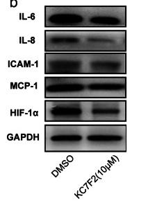
Application: WB    Species: mouse    Sample: primary bone marrow endothelial cells

Fig. 4 |Characterization of primary bone marrow endothelial cells by RT-QPCR and immunoblottinge .The immunoblotting analysis of primary bone marrow endothelial after 7 days of cultured further substantiates the purity of endothelial cells with protein ladder (M) (N= 3 indicates independent experiments for RT-qPCR and N=2 for immunoblotting with six mice/group(12 femora and 12 tibias). *p<0.05 **p<0.01, and ***p<0.001

Application: IF/ICC    Species: mouse    Sample: primary bone marrow endothelial cells

Fig. 7| a, b Bone marrow endothelial cell proliferation Ki-67 by flow cytometry analysis and hemocytometer.e The merged immunofluorescence staining of both quiescence BMECs and treated endothelial cells with TNF-α. Scale bar = 5μm, ×20 magnification, N= 3 experimental repeats 3 mice per group, *p< 0.05, **p<0.01, and ***p<0.001

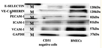
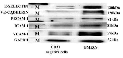
Application: WB    Species: Mice    Sample: bMECs

Fig. 4 Characterization of primary bone marrow endothelial cells by RT-QPCR and immunoblotting: a–d the relative mRNA expression of primary bone marrow endothelial cells compared to CD31 microbead-negative selected cells. The bar chart indicated the fold of the gene expression of primary BMECs compared to the CD31 microbead-negative selected cells. The fold change is quantified by the Pitfall method, followed by a Student’s test comparison between the CD31 microbead-negative selected cells and endothelial cells. P value ≤0.05 considered being statistically significant. e The immunoblotting analysis of primary bone marrow endothelial after 7 days of cultured further substantiates the purity of endothelial cells with protein ladder (M) (N= 3 indicates independent experiments for RT-qPCR and N=2 for immunoblotting with six mice/group (12 femora and 12 tibias). *p<0.05 **p<0.01, and ***p<0.001

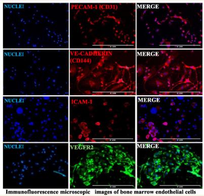
Application: IF/ICC    Species: Mice    Sample: BMECs

Fig. 5 Identification of primary bone marrow endothelial cells by immunofluorescence staining: a anti-CD31 (PECAM-1), anti-CD144 (VE-cadherin), anti-CD106 (ICAM-1), and anti-VEGFR2—the cell shows the expression of these endothelial cell-specific molecules. b The mean fluorescence intensity for endothelial cells particular marker in the graph was relatively quantified by ImageJ, which shows the order of the endothelial cell-specific markers (PECAM-1> VE-cadherin > ICAM-1 > VEGFR2, N = 3 experimental repeats with three mice/group (6 femora and 6 tibias), scale bar 5μm, ×20 magnification

4). The reduced SCFA-producing gut microbes are involved in the inflammatory activation in Kawasaki disease. Frontiers in Immunology (PubMed: 37398673) [IF=7.3]

Application: IF/ICC    Species: Mouse    Sample:

Figure 1 Gut dysbiosis played a role in the inflammation and coronary artery injury of the KD mouse model. (A) PCoA plots of bray-curtis similarity matrices between PBS group (n=4) and CAWS group (n=7). (B) Heatmap of gut microbiota on the genus level. (C) The relative abundance of the gut microbiota in phylum level. (D) Plasma cytokine levels of IL-1β, IL-6, TNF-α, MCP-1. (E) H&E staining of heart tissues. The coronary artery lesion in the CAWS group was circled in red and pointed out by an arrow. Magnification: ×200. Scale bar = 20 μm. (F, G) The expression levels of intercellular cell adhesion molecule-1 (ICAM-1) and macrophage marker CD68 were evaluated using immunofluorescence of coronary arteries. Magnification: × 200. Scale bars = 20 µm. Values are expressed as the mean ± SEM. Significance: ** P < 0.01, *** P < 0.001, vs. the PBS group; # P < 0.05, ## P < 0.01 vs. the CAWS group; ns means no significance.

5). CXCR1 and its downstream NF-κB inflammation signaling pathway as a key target of Guanxinning injection for myocardial ischemia/reperfusion injury. Frontiers in Immunology (PubMed: 36325326) [IF=7.3]

6). Karyopherin α 2 promotes proliferation, migration and invasion through activating NF-κB/p65 signaling pathways in melanoma cells. LIFE SCIENCES (PubMed: 32243925) [IF=6.1]

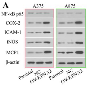
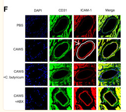
Application: WB    Species: Mice    Sample: melanoma cells

Fig. 4. KPNA2 activated NF-κB/p65 signaling pathway in melanoma cells. The expression of NF-κB p65, COX-2, ICAM-1, iNOS, MCP1, and p65 (nucleus) was analyzed by western blot in the KPNA2-overexpressing (A, B) or KPNA2-silenced cells (F, G). The nuclear translocation of p65 (nucleus) was detected by immunofluorescence assay in the KPNA2-overexpressing (C, D) or KPNA2-silenced cells (H, I). The NF-κB binding activity was analyzed by EMSA in the KPNA2- overexpressing (E) or KPNA2-silenced cells (J). Parental, blank control group; NC, negative control group; OV-KPNA2, KPNA2 overexpressed group; siRNA1-KPNA2, KPNA2-1 silencing group; siRNA2-KPNA2, KPNA2-2 silencing group. The results were obtained in three independent experiments. Mean values were compared by One-way ANOVA. (**p < 0.01, ***p < 0.001, ****p < 0.001).

7). Resolvin D1 ameliorates Inflammation-Mediated Blood-Brain Barrier Disruption After Subarachnoid Hemorrhage in rats by Modulating A20 and NLRP3 Inflammasome. Frontiers in Pharmacology (PubMed: 33732145) [IF=5.6]

8). Zinc attenuates ferroptosis and promotes functional recovery in contusion spinal cord injury by activating Nrf2/GPX4 defense pathway. CNS Neuroscience & Therapeutics (PubMed: 33951302) [IF=5.5]

Application: WB    Species: Mouse    Sample: Spinal cord

FIGURE 7 Zinc can reduce the inflammation of damaged parts by inhibiting ferroptosis. (A) The expressions of TNF-α, IL-6, IL-1β, and ICAM-1 were evaluated by Western blotting at 3 days post-SCI in each group (n = 6). (B) Quantification of TNF-α, IL-6, IL-1β, and ICAM-1 expressions (n = 6, all the data are expressed as means ± SD, two-way ANOVA followed by Tukey's post hoc test was applied). (C–F) Immunofluorescence staining was used to detect the level of TNF-α, IL-6, IL-1β, and ICAM-1 from each group (n = 6, scale bar = 50 µm). (G) Statistical analysis of immunofluorescence staining for positive expression of TNF-α, IL-6, IL-1β, and ICAM-1 in nerve cells from each group (n = 6, all the data are expressed as means ± SD, two-way ANOVA followed by Tukey's post hoc test was applied). * means p < 0.05; **means p < 0.01; and *** means p < 0.001

9). Nepeta angustifolia attenuates responses to vascular inflammation in high glucose-induced human umbilical vein endothelial cells through heme oxygenase-1 induction. JOURNAL OF ETHNOPHARMACOLOGY (PubMed: 30419276) [IF=5.4]

10). Sanhuang Xiexin decoction ameliorates DSS-induced colitis in mice by regulating intestinal inflammation, intestinal barrier, and intestinal flora. JOURNAL OF ETHNOPHARMACOLOGY (PubMed: 35843414) [IF=5.4]

Load more

Restrictive clause


Affinity Biosciences tests all products strictly. Citations are provided as a resource for additional applications that have not been validated by Affinity Biosciences. Please choose the appropriate format for each application and consult Materials and Methods sections for additional details about the use of any product in these publications.

For Research Use Only.
Not for use in diagnostic or therapeutic procedures. Not for resale. Not for distribution without written consent. Affinity Biosciences will not be held responsible for patent infringement or other violations that may occur with the use of our products. Affinity Biosciences, Affinity Biosciences Logo and all other trademarks are the property of Affinity Biosciences LTD.