 antibody.")
 The mRNA levels of FUNDC1, LC3A, LC3B and PGAM5. (B) The western blot results of FUNDC1, p-FUNDC1 and LC3. (C) The quantification of FUNDC1, p-FUNDC1 (Ser17) and LC3II/LC3I protein levels. (D) Immunofluorescence co-location of COX IV and LC3 at day 50. In the images, the nucleus staining is shown in blue, COX IV staining is shown in green, LC3 staining is shown in red, and the signals of colocalization are shown in merged images. (E) Pearson coefficient of the colocalization of COX IV and LC3. Data are expressed as means ± SD (n = 6). “*” indicates significant difference compared to the corresponding control (*P < 0.05, **P < 0.01). “#” indicates statistically significant difference between corresponding groups (#P < 0.05, ##P < 0.01).")
 The expression levels of ULK1, PGAM5, FUNDC1, p-FUNDC1 and GAPDH. (B) ULK1 relative to GAPDH, (C) PGAM5 relative to GAPDH, (D) FUNDC1 relative to GAPDH. (E) p-FUNDC1 relative to GAPDH. (F) ULK mRNA levels. (G) PGAM5 mRNA levels. (H) FUNDC1 mRNA levels. **P<0.05 vs. C group and #P<0.05 vs. DOX group. Protein levels were measured by western blotting. ULK1, PGAM5 and FUNDC1 mRNA levels were measured by RT-qPCR. Data are mean ± standard deviation from five independent experiments (n=13-20). MOX moxibustion; FUNDC1, FUN14 domain-containing protein 1; ULK1, Unc-51 Like Autophagy Activating Kinase 1; PGAM5, phosphoglycerate mutase family member 5; p-, phosphorylated; DOX, doxorubicin; BEN, benazepril C, control.")
 mRNA expression of IL-1β, IL-6, IL-8 and TNF-α in human healthy and pulpitis tissues. mRNA expression of (B) HIF-1α and (C) FUNDC1 in human healthy and pulpitis tissues. (D) Representative immunostaining images of HIF-1α and FUNDC1 in human healthy or inflamed dental pulp tissues. Scale bars are 100 and 25 µm, respectively. Results are presented as the means ± SD from ≥ three independent experiments. *P<0.05, **P<0.01 and ***P<0.001 vs. healthy. HIF-1α, hypoxia-inducible factor-1α; FUNDC1, FUN14 domain-containing 1.")
. The serum AMH (B), E2 (C), FSH (D), and LH (E) levels. F Ki67 expression in ovarian GCs; scale bar: 100 μm. G The Western blotting images and quantitation of functional protein expression in GCs. H The fertility and litter size.")
| Product: | Phospho-FUNDC1 (Ser17) antibody |
| Catalog: | AF0001 |
| Description: | Rabbit polyclonal antibody to Phospho-FUNDC1 (Ser17) |
| Application: | WB |
| Cited expt.: | WB |
| Reactivity: | Human |
| Prediction: | Pig, Bovine, Horse, Sheep, Rabbit, Dog, Chicken |
| Mol.Wt.: | 17KD(Observed); 17kD(Calculated). |
| Uniprot: | Q8IVP5 |
| RRID: | AB_2846773 |
Control Products

Product Info
*The optimal dilutions should be determined by the end user. For optimal experimental results, antibody reuse is not recommended.
*Tips:
WB: For western blot detection of denatured protein samples. IHC: For immunohistochemical detection of paraffin sections (IHC-p) or frozen sections (IHC-f) of tissue samples. IF/ICC: For immunofluorescence detection of cell samples. ELISA(peptide): For ELISA detection of antigenic peptide.
Cite Format: Affinity Biosciences Cat# AF0001, RRID:AB_2846773.
Fold/Unfold
FUN14 domain containing 1; FUN14 domain containing protein 1; FUN14 domain-containing protein 1; FUND1_HUMAN; fundc1;
Immunogens
A synthesized peptide derived from human FUNDC1 around the phosphorylation site of Serine 17.
- Q8IVP5 FUND1_HUMAN:
- Protein BLAST With
- NCBI/
- ExPASy/
- Uniprot
MATRNPPPQDYESDDDSYEVLDLTEYARRHQWWNRVFGHSSGPMVEKYSVATQIVMGGVTGWCAGFLFQKVGKLAATAVGGGFLLLQIASHSGYVQIDWKRVEKDVNKAKRQIKKRANKAAPEINNLIEEATEFIKQNIVISSGFVGGFLLGLAS
Predictions
Score>80(red) has high confidence and is suggested to be used for WB detection. *The prediction model is mainly based on the alignment of immunogen sequences, the results are for reference only, not as the basis of quality assurance.
High(score>80) Medium(80>score>50) Low(score<50) No confidence
Research Backgrounds
Acts as an activator of hypoxia-induced mitophagy, an important mechanism for mitochondrial quality control.
Phosphorylation at Tyr-18 by SRC inhibits activation of mitophagy. Following hypoxia, dephosphorylated at Tyr-18, leading to interaction with MAP1 LC3 family proteins and triggering mitophagy.
Mitochondrion outer membrane>Multi-pass membrane protein.
Widely expressed.
The YXXL motif mediates the interaction with MAP1 LC3 family proteins MAP1LC3A, MAP1LC3B and GABARAP.
Belongs to the FUN14 family.
References

Application: WB Species: Mouse Sample:

Application: WB Species: rat Sample:
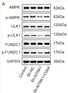
Application: WB Species: Mouse Sample:
Restrictive clause
Affinity Biosciences tests all products strictly. Citations are provided as a resource for additional applications that have not been validated by Affinity Biosciences. Please choose the appropriate format for each application and consult Materials and Methods sections for additional details about the use of any product in these publications.
For Research Use Only.
Not for use in diagnostic or therapeutic procedures. Not for resale. Not for distribution without written consent. Affinity Biosciences will not be held responsible for patent infringement or other violations that may occur with the use of our products. Affinity Biosciences, Affinity Biosciences Logo and all other trademarks are the property of Affinity Biosciences LTD.