CD20 Antibody - #DF13319

Related Downloads
Protocols
Product Info
*The optimal dilutions should be determined by the end user.
*Tips:
WB: For western blot detection of denatured protein samples. IHC: For immunohistochemical detection of paraffin sections (IHC-p) or frozen sections (IHC-f) of tissue samples. IF/ICC: For immunofluorescence detection of cell samples. ELISA(peptide): For ELISA detection of antigenic peptide.
Cite Format: Affinity Biosciences Cat# DF13319, RRID:AB_2846338.
Fold/Unfold
APY; ATOPY; B lymphocyte antigen CD20; B Lymphocyte Cell Surface Antigen B1; B-lymphocyte antigen CD20; B-lymphocyte cell-surface antigen B1; B-lymphocyte surface antigen B1; B1; Bp 35; Bp35; CD 20; CD20; CD20 antigen; CD20 receptor; CD20_HUMAN; CVID5; Fc epsilon receptor I beta chain; Fc Fragment of IgE high affinity I receptor for beta polypeptide; FCER1B; High affinity immunoglobulin epsilon receptor subunit beta; IgE Fc receptor subunit beta; IGEL; IGER; IGHER; LEU 16; LEU16; leukocyte surface antigen Leu 16; Leukocyte surface antigen Leu-16; Ly44; Membrane spanning 4 domains A1; Membrane spanning 4 domains subfamily A member 2; Membrane-spanning 4-domains subfamily A member 1; MGC3969; MS4A1; MS4A2; S7;
Immunogens
- P11836 CD20_HUMAN:
- Protein BLAST With
- NCBI/
- ExPASy/
- Uniprot
MTTPRNSVNGTFPAEPMKGPIAMQSGPKPLFRRMSSLVGPTQSFFMRESKTLGAVQIMNGLFHIALGGLLMIPAGIYAPICVTVWYPLWGGIMYIISGSLLAATEKNSRKCLVKGKMIMNSLSLFAAISGMILSIMDILNIKISHFLKMESLNFIRAHTPYINIYNCEPANPSEKNSPSTQYCYSIQSLFLGILSVMLIFAFFQELVIAGIVENEWKRTCSRPKSNIVLLSAEEKKEQTIEIKEEVVGLTETSSQPKNEEDIEIIPIQEEEEEETETNFPEPPQDQESSPIENDSSP
Predictions
Score>80(red) has high confidence and is suggested to be used for WB detection. *The prediction model is mainly based on the alignment of immunogen sequences, the results are for reference only, not as the basis of quality assurance.
High(score>80) Medium(80>score>50) Low(score<50) No confidence
PTMs - P11836 As Substrate
| Site | PTM Type | Enzyme | Source |
|---|---|---|---|
| T2 | Phosphorylation | Uniprot | |
| T3 | Phosphorylation | Uniprot | |
| S7 | Phosphorylation | Uniprot | |
| T11 | Phosphorylation | Uniprot | |
| S25 | Phosphorylation | Uniprot | |
| S35 | Phosphorylation | Uniprot | |
| S36 | Phosphorylation | Uniprot | |
| T41 | Phosphorylation | Uniprot | |
| S121 | Phosphorylation | Uniprot | |
| S225 | Phosphorylation | Uniprot | |
| S231 | Phosphorylation | P68400 (CSNK2A1) , P19784 (CSNK2A2) | Uniprot |
| K235 | Ubiquitination | Uniprot | |
| K236 | Ubiquitination | Uniprot | |
| K243 | Ubiquitination | Uniprot | |
| T250 | Phosphorylation | P68400 (CSNK2A1) , P19784 (CSNK2A2) | Uniprot |
| S253 | Phosphorylation | Uniprot | |
| S254 | Phosphorylation | Uniprot | |
| S289 | Phosphorylation | P68400 (CSNK2A1) , P19784 (CSNK2A2) | Uniprot |
Research Backgrounds
B-lymphocyte-specific membrane protein that plays a role in the regulation of cellular calcium influx necessary for the development, differentiation, and activation of B-lymphocytes. Functions as a store-operated calcium (SOC) channel component promoting calcium influx after activation by the B-cell receptor/BCR.
Phosphorylated on serines and threonines in resting B-cells. Protein kinase C/PKC can use CD20 as substrate.
Cell membrane>Multi-pass membrane protein. Cell membrane>Lipid-anchor.
Note: Constitutively associated with membrane rafts.
Expressed on B-cells.
Forms homotetramers. Interacts with the heavy and light chains of cell surface IgM, the antigen-binding components of the BCR.
Belongs to the MS4A family.
Research Fields
· Organismal Systems > Immune system > Hematopoietic cell lineage. (View pathway)
References
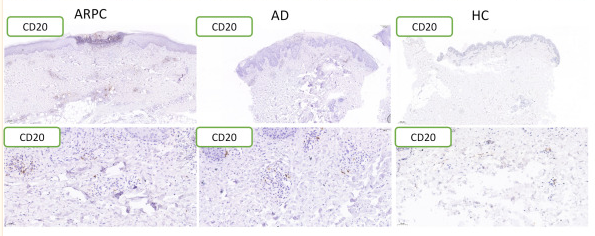
Application: IHC Species: Human Sample:
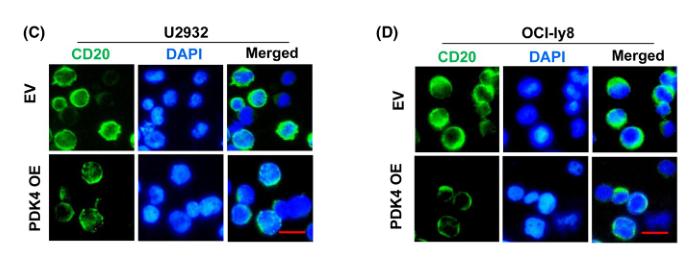
Application: IF/ICC Species: Human Sample: DLBCL cells

Application: WB Species: Human Sample: DLBCL cells
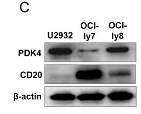
Application: WB Species: Human Sample: DLBCL cells
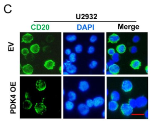
Application: IF/ICC Species: Human Sample: DLBCL cells
Restrictive clause
Affinity Biosciences tests all products strictly. Citations are provided as a resource for additional applications that have not been validated by Affinity Biosciences. Please choose the appropriate format for each application and consult Materials and Methods sections for additional details about the use of any product in these publications.
For Research Use Only.
Not for use in diagnostic or therapeutic procedures. Not for resale. Not for distribution without written consent. Affinity Biosciences will not be held responsible for patent infringement or other violations that may occur with the use of our products. Affinity Biosciences, Affinity Biosciences Logo and all other trademarks are the property of Affinity Biosciences LTD.