CD36 Antibody - #DF13262

Related Downloads
Protocols
Product Info
*The optimal dilutions should be determined by the end user.
*Tips:
WB: For western blot detection of denatured protein samples. IHC: For immunohistochemical detection of paraffin sections (IHC-p) or frozen sections (IHC-f) of tissue samples. IF/ICC: For immunofluorescence detection of cell samples. ELISA(peptide): For ELISA detection of antigenic peptide.
Cite Format: Affinity Biosciences Cat# DF13262, RRID:AB_2846281.
Fold/Unfold
Adipocyte membrane protein; BDPLT10; CD36; CD36 antigen (collagen type I receptor, thrombospondin receptor); CD36 antigen; CD36 molecule (thrombospondin receptor); CD36 molecule; CD36_HUMAN; CHDS7; Cluster determinant 36; Collagen receptor, platelet; FAT; Fatty acid translocase; Fatty acid transport protein; Glycoprotein IIIb; GP IIIb; GP3B; GP4; GPIIIB; GPIV; Leukocyte differentiation antigen CD36; MGC108510; MGC91634; PAS 4 protein; PAS IV; PAS-4; PASIV; Platelet collagen receptor; Platelet glycoprotein 4; Platelet glycoprotein IV; scarb3; Scavenger receptor class B member 3; Thrombospondin receptor;
Immunogens
- P16671 CD36_HUMAN:
- Protein BLAST With
- NCBI/
- ExPASy/
- Uniprot
MGCDRNCGLIAGAVIGAVLAVFGGILMPVGDLLIQKTIKKQVVLEEGTIAFKNWVKTGTEVYRQFWIFDVQNPQEVMMNSSNIQVKQRGPYTYRVRFLAKENVTQDAEDNTVSFLQPNGAIFEPSLSVGTEADNFTVLNLAVAAASHIYQNQFVQMILNSLINKSKSSMFQVRTLRELLWGYRDPFLSLVPYPVTTTVGLFYPYNNTADGVYKVFNGKDNISKVAIIDTYKGKRNLSYWESHCDMINGTDAASFPPFVEKSQVLQFFSSDICRSIYAVFESDVNLKGIPVYRFVLPSKAFASPVENPDNYCFCTEKIISKNCTSYGVLDISKCKEGRPVYISLPHFLYASPDVSEPIDGLNPNEEEHRTYLDIEPITGFTLQFAKRLQVNLLVKPSEKIQVLKNLKRNYIVPILWLNETGTIGDEKANMFRSQVTGKINLLGLIEMILLSVGVVMFVAFMISYCACRSKTIK
Predictions
Score>80(red) has high confidence and is suggested to be used for WB detection. *The prediction model is mainly based on the alignment of immunogen sequences, the results are for reference only, not as the basis of quality assurance.
High(score>80) Medium(80>score>50) Low(score<50) No confidence
PTMs - P16671 As Substrate
| Site | PTM Type | Enzyme | Source |
|---|---|---|---|
| T57 | Phosphorylation | Uniprot | |
| Y62 | Phosphorylation | Uniprot | |
| T92 | Phosphorylation | P17252 (PRKCA) | Uniprot |
| N205 | N-Glycosylation | Uniprot | |
| K298 | Acetylation | Uniprot | |
| N321 | N-Glycosylation | Uniprot | |
| T323 | Phosphorylation | Uniprot | |
| N417 | N-Glycosylation | Uniprot | |
| K469 | Ubiquitination | Uniprot | |
| K472 | Ubiquitination | Uniprot |
Research Backgrounds
Multifunctional glycoprotein that acts as receptor for a broad range of ligands. Ligands can be of proteinaceous nature like thrombospondin, fibronectin, collagen or amyloid-beta as well as of lipidic nature such as oxidized low-density lipoprotein (oxLDL), anionic phospholipids, long-chain fatty acids and bacterial diacylated lipopeptides. They are generally multivalent and can therefore engage multiple receptors simultaneously, the resulting formation of CD36 clusters initiates signal transduction and internalization of receptor-ligand complexes. The dependency on coreceptor signaling is strongly ligand specific. Cellular responses to these ligands are involved in angiogenesis, inflammatory response, fatty acid metabolism, taste and dietary fat processing in the intestine (Probable). Binds long-chain fatty acids and facilitates their transport into cells, thus participating in muscle lipid utilization, adipose energy storage, and gut fat absorption (By similarity). In the small intestine, plays a role in proximal absorption of dietary fatty acid and cholesterol for optimal chylomicron formation, possibly through the activation of MAPK1/3 (ERK1/2) signaling pathway (By similarity). Involved in oral fat perception and preferences. Detection into the tongue of long-chain fatty acids leads to a rapid and sustained rise in flux and protein content of pancreatobiliary secretions (By similarity). In taste receptor cells, mediates the induction of an increase in intracellular calcium levels by long-chain fatty acids, leading to the activation of the gustatory neurons in the nucleus of the solitary tract (By similarity). Important factor in both ventromedial hypothalamus neuronal sensing of long-chain fatty acid and the regulation of energy and glucose homeostasis (By similarity). Receptor for thombospondins, THBS1 and THBS2, mediating their antiangiogenic effects (By similarity). As a coreceptor for TLR4:TLR6 heterodimer, promotes inflammation in monocytes/macrophages. Upon ligand binding, such as oxLDL or amyloid-beta 42, interacts with the heterodimer TLR4:TLR6, the complex is internalized and triggers inflammatory response, leading to NF-kappa-B-dependent production of CXCL1, CXCL2 and CCL9 cytokines, via MYD88 signaling pathway, and CCL5 cytokine, via TICAM1 signaling pathway, as well as IL1B secretion, through the priming and activation of the NLRP3 inflammasome (By similarity). Selective and nonredundant sensor of microbial diacylated lipopeptide that signal via TLR2:TLR6 heterodimer, this cluster triggers signaling from the cell surface, leading to the NF-kappa-B-dependent production of TNF, via MYD88 signaling pathway and subsequently is targeted to the Golgi in a lipid-raft dependent pathway (By similarity).
(Microbial infection) Directly mediates cytoadherence of Plasmodium falciparum parasitized erythrocytes and the internalization of particles independently of TLR signaling.
N-glycosylated and O-glycosylated with a ratio of 2:1.
Ubiquitinated at Lys-469 and Lys-472. Ubiquitination is induced by fatty acids such as oleic acid and leads to degradation by the proteasome. Ubiquitination and degradation are inhibited by insulin which blocks the effect of fatty acids.
Cell membrane>Multi-pass membrane protein. Membrane raft. Golgi apparatus. Apical cell membrane.
Note: Upon ligand-binding, internalized through dynamin-dependent endocytosis.
Interacts with THBS1 and THBS2; the interactions mediate the THBS antiangiogenic activity. Upon interaction with a ligand, such as oxidized low-density lipoprotein (oxLDL) or amyloid-beta 42, rapidly forms a complex with TLR4 and TLR6; the complex is internalized and triggers an inflammatory signal. Through its C-terminus, interacts with PTK2, PXN and LYN, but not with SRC. LYN kinase activity is required for facilitating TLR4:TLR6 heterodimerization and signal initiation. Upon interaction with ligands such as diacylated lipopeptides, interacts with the TLR2:TLR6 heterodimer. Interacts with CD9, CD81, FCER1G, ITGB2 and/or ITGB2; forming a membrane heteromeric complex required for the internalization of CD36 and its ligands (By similarity).
(Microbial infection) Binds to Plasmodium falciparum EMP1.
Belongs to the CD36 family.
Research Fields
· Cellular Processes > Transport and catabolism > Phagosome. (View pathway)
· Environmental Information Processing > Signal transduction > AMPK signaling pathway. (View pathway)
· Environmental Information Processing > Signaling molecules and interaction > ECM-receptor interaction. (View pathway)
· Human Diseases > Endocrine and metabolic diseases > Insulin resistance.
· Human Diseases > Infectious diseases: Parasitic > Malaria.
· Organismal Systems > Endocrine system > PPAR signaling pathway.
· Organismal Systems > Immune system > Hematopoietic cell lineage. (View pathway)
· Organismal Systems > Endocrine system > Adipocytokine signaling pathway.
· Organismal Systems > Digestive system > Fat digestion and absorption.
· Organismal Systems > Digestive system > Cholesterol metabolism.
References

Application: WB Species: Mouse Sample: OE APOC2 AGS cells
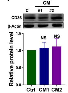
Application: WB Species: human Sample: DLD1 cells
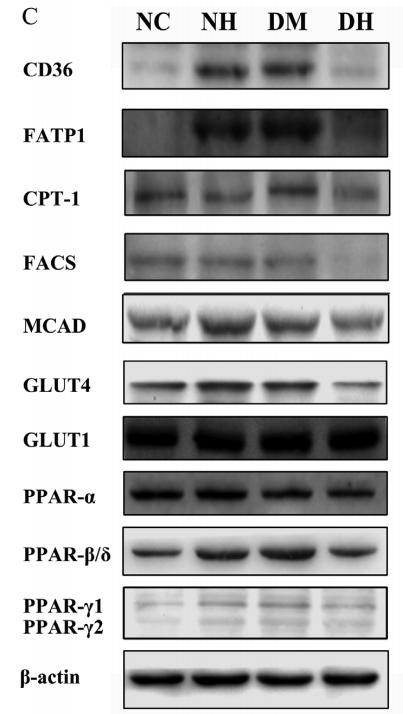
Application: WB Species: mouse Sample: myocardial
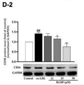
Application: WB Species: Mice Sample: RAW264.7 cells
Restrictive clause
Affinity Biosciences tests all products strictly. Citations are provided as a resource for additional applications that have not been validated by Affinity Biosciences. Please choose the appropriate format for each application and consult Materials and Methods sections for additional details about the use of any product in these publications.
For Research Use Only.
Not for use in diagnostic or therapeutic procedures. Not for resale. Not for distribution without written consent. Affinity Biosciences will not be held responsible for patent infringement or other violations that may occur with the use of our products. Affinity Biosciences, Affinity Biosciences Logo and all other trademarks are the property of Affinity Biosciences LTD.