Product: CX3CL1 Antibody
Catalog: DF12376
Description: Rabbit polyclonal antibody to CX3CL1
Application: WB IHC IF/ICC
Reactivity: Human, Mouse
Mol.Wt.: 90-100 kDa; 42kD(Calculated).
Uniprot: P78423
RRID: AB_2845181

View similar products>>

   Size Price Inventory
 100ul $280 In stock
 200ul $350 In stock

Lead Time: Same day delivery

For pricing and ordering contact:
Local distributors

Product Info

Source:
Rabbit
Application:
WB 1:500-1:2000, IHC 1:50-1:200, IF/ICC 1:100-1:500
*The optimal dilutions should be determined by the end user.
*Tips:

WB: For western blot detection of denatured protein samples. IHC: For immunohistochemical detection of paraffin sections (IHC-p) or frozen sections (IHC-f) of tissue samples. IF/ICC: For immunofluorescence detection of cell samples. ELISA(peptide): For ELISA detection of antigenic peptide.

Reactivity:
Human,Mouse
Clonality:
Polyclonal
Specificity:
CX3CL1 Antibody detects endogenous levels of total CX3CL1.
RRID:
AB_2845181
Cite Format: Affinity Biosciences Cat# DF12376, RRID:AB_2845181.
Conjugate:
Unconjugated.
Purification:
The antiserum was purified by peptide affinity chromatography using SulfoLink™ Coupling Resin (Thermo Fisher Scientific).
Storage:
Rabbit IgG in phosphate buffered saline , pH 7.4, 150mM NaCl, 0.02% sodium azide and 50% glycerol. Store at -20 °C. Stable for 12 months from date of receipt.
Alias:

Fold/Unfold

A 152E5.2; AB030188; ABCD 3; ABCD3; AI848747; C-X3-C motif chemokine 1; C3Xkine; Chemokine (C-X3-C motif) ligand 1; Chemokine C X3 C motif ligand 1; Chemokine CX3C Motif Ligand 1; CX3C membrane anchored chemokine; CX3C membrane-anchored chemokine; Cx3cl1; CXC 3; CXC3; CXC3C; D8Bwg0439e; FKN; Fractalkine; Neurotactin; NTN; NTT; Processed fractalkine; SCYD 1; SCYD1; Small inducible cytokine D1; Small inducible cytokine subfamily D (Cys X3 Cys) member 1; small inducible cytokine subfamily D (Cys-X3-Cys), member 1 (fractalkine, neurotactin); Small inducible cytokine subfamily D member 1; Small-inducible cytokine D1; X3CL1_HUMAN;

Immunogens

Immunogen:
Uniprot:
Gene(ID):
Expression:
P78423 X3CL1_HUMAN:

Expressed in the seminal plasma, endometrial fluid and follicular fluid (at protein level). Small intestine, colon, testis, prostate, heart, brain, lung, skeletal muscle, kidney and pancreas. Most abundant in the brain and heart.

Sequence:
MAPISLSWLLRLATFCHLTVLLAGQHHGVTKCNITCSKMTSKIPVALLIHYQQNQASCGKRAIILETRQHRLFCADPKEQWVKDAMQHLDRQAAALTRNGGTFEKQIGEVKPRTTPAAGGMDESVVLEPEATGESSSLEPTPSSQEAQRALGTSPELPTGVTGSSGTRLPPTPKAQDGGPVGTELFRVPPVSTAATWQSSAPHQPGPSLWAEAKTSEAPSTQDPSTQASTASSPAPEENAPSEGQRVWGQGQSPRPENSLEREEMGPVPAHTDAFQDWGPGSMAHVSVVPVSSEGTPSREPVASGSWTPKAEEPIHATMDPQRLGVLITPVPDAQAATRRQAVGLLAFLGLLFCLGVAMFTYQSLQGCPRKMAGEMAEGLRYIPRSCGSNSYVLVPV

PTMs - P78423 As Substrate

Site PTM Type Enzyme
T97 Phosphorylation
T183 O-Glycosylation
S225 Phosphorylation
S253 O-Glycosylation
T318 O-Glycosylation
T329 O-Glycosylation
S391 Phosphorylation

Research Backgrounds

Function:

Acts as a ligand for both CX3CR1 and integrins. Binds to CX3CR1. Binds to integrins ITGAV:ITGB3 and ITGA4:ITGB1. Can activate integrins in both a CX3CR1-dependent and CX3CR1-independent manner. In the presence of CX3CR1, activates integrins by binding to the classical ligand-binding site (site 1) in integrins. In the absence of CX3CR1, binds to a second site (site 2) in integrins which is distinct from site 1 and enhances the binding of other integrin ligands to site 1. The soluble form is chemotactic for T-cells and monocytes and not for neutrophils. The membrane-bound form promotes adhesion of those leukocytes to endothelial cells. May play a role in regulating leukocyte adhesion and migration processes at the endothelium.

PTMs:

A soluble short 95 kDa form may be released by proteolytic cleavage from the long membrane-anchored form.

O-glycosylated with core 1 or possibly core 8 glycans.

Subcellular Location:

Cell membrane>Single-pass type I membrane protein.

Secreted.

Extracellular region or secreted Cytosol Plasma membrane Cytoskeleton Lysosome Endosome Peroxisome ER Golgi apparatus Nucleus Mitochondrion Manual annotation Automatic computational assertionSubcellular location
Tissue Specificity:

Expressed in the seminal plasma, endometrial fluid and follicular fluid (at protein level). Small intestine, colon, testis, prostate, heart, brain, lung, skeletal muscle, kidney and pancreas. Most abundant in the brain and heart.

Subunit Structure:

Monomer. Interacts with pox virus crmD; this inhibits cell migration mediated by CX3CL1. Forms a ternary complex with CX3CR1 and ITGAV:ITGB3 or ITGA4:ITGB1.

Family&Domains:

Belongs to the intercrine delta family.

Research Fields

· Environmental Information Processing > Signaling molecules and interaction > Cytokine-cytokine receptor interaction.   (View pathway)

· Environmental Information Processing > Signal transduction > TNF signaling pathway.   (View pathway)

· Organismal Systems > Immune system > Chemokine signaling pathway.   (View pathway)

References

1). Identification of Ligand-Receptor Pairs Associated With Tumour Characteristics in Clear Cell Renal Cell Carcinoma. Frontiers in Immunology (PubMed: 35734169) [IF=7.3]

Application: WB    Species: Human    Sample: ccRCC cell line 786-O and renal epithelial cel

Figure 12 CX3CL1 Knockdown promoted migration and invasion of ccRCC cell in vitro. (A) Real-time RT-PCR assay of 10 LR-pairs mRNA expression levels in human ccRCC cell line 786-O and renal epithelial cell HK-2. (B, C) the protein expression of CX3CL1 was performed by Western Blot assay. (D) The subcellular localization of CX3CL1 was identified by immunostaining assay. Scale bar: 10μm. (E) Cell colonies of 786-O cell in normal control and CX3CL1-KD group. (F, G) 786-O cell migration assay (F) and invasion assay (G) were performed in normal control and CX3CL1-KD group. ns, p≥0.05; *p< 0.05; **p<0.01; ***p<0.001.

Application: IF/ICC    Species: Human    Sample: ccRCC cell line 786-O and renal epithelial cel

Figure 12 CX3CL1 Knockdown promoted migration and invasion of ccRCC cell in vitro. (A) Real-time RT-PCR assay of 10 LR-pairs mRNA expression levels in human ccRCC cell line 786-O and renal epithelial cell HK-2. (B, C) the protein expression of CX3CL1 was performed by Western Blot assay. (D) The subcellular localization of CX3CL1 was identified by immunostaining assay. Scale bar: 10μm. (E) Cell colonies of 786-O cell in normal control and CX3CL1-KD group. (F, G) 786-O cell migration assay (F) and invasion assay (G) were performed in normal control and CX3CL1-KD group. ns, p≥0.05; *p< 0.05; **p<0.01; ***p<0.001.

Application: IHC    Species: Human    Sample: ccRCC cell line 786-O and renal epithelial cel

Figure 12 CX3CL1 Knockdown promoted migration and invasion of ccRCC cell in vitro. (A) Real-time RT-PCR assay of 10 LR-pairs mRNA expression levels in human ccRCC cell line 786-O and renal epithelial cell HK-2. (B, C) the protein expression of CX3CL1 was performed by Western Blot assay. (D) The subcellular localization of CX3CL1 was identified by immunostaining assay. Scale bar: 10μm. (E) Cell colonies of 786-O cell in normal control and CX3CL1-KD group. (F, G) 786-O cell migration assay (F) and invasion assay (G) were performed in normal control and CX3CL1-KD group. ns, p≥0.05; *p< 0.05; **p<0.01; ***p<0.001.

2). Platelets are recruited to hepatocellular carcinoma tissues in a CX3CL1‐CX3CR1 dependent manner and induce tumour cell apoptosis. Molecular Oncology (PubMed: 32799418) [IF=6.6]

Application: IHC    Species: human    Sample: liver

Fig. 4. |Hypoxia promotes platelet recruitment by upregulating CX3CL1 expression.(C) Expression of CX3CL1 and CA9 in human HCC and NT liver tissues. Representative images of two consecutive slices of the same sample embedded in paraffin were taken, and each was stained with CX3CL1 and CA9 (n = 3). Bar, 200 lm.

3). Fractalkine Aggravates LPS-induced Macrophage Activation and Acute Kidney Injury via Wnt/β-catenin Signaling Pathway. JOURNAL OF CELLULAR AND MOLECULAR MEDICINE (PubMed: 34101346) [IF=5.3]

Application: WB    Species: Mice    Sample: J774A.1 cells

FIGURE 1 FKN positively interacted with Wnt/β- catenin signalling pathway in J774A.1 cells. A, Expression profile of FKN- regulated genes using the RNA- seq. B, Differentially expressed genes are shown in a volcano map. C, Biological processes were revealed in RNA- sequencing. D, The data sets were analysed by GESA using the Hallmark collection. The correlation between FKN and Wnt/β- catenin signalling was indicated in GESA. E- G, Co- IP and IF assays detected the interaction between FKN and Wnt/β- catenin signalling and this was visualized using confocal microscopy with specific antibodies (green). Nuclei were incubated with DAPI (blue). The cytoskeletons were incubated with β- tubulin (red). Scale bars represent 10 μm

Application: IF/ICC    Species: Mice    Sample: J774A.1 cells

FIGURE 1 FKN positively interacted with Wnt/β- catenin signalling pathway in J774A.1 cells. A, Expression profile of FKN- regulated genes using the RNA- seq. B, Differentially expressed genes are shown in a volcano map. C, Biological processes were revealed in RNA- sequencing. D, The data sets were analysed by GESA using the Hallmark collection. The correlation between FKN and Wnt/β- catenin signalling was indicated in GESA. E- G, Co- IP and IF assays detected the interaction between FKN and Wnt/β- catenin signalling and this was visualized using confocal microscopy with specific antibodies (green). Nuclei were incubated with DAPI (blue). The cytoskeletons were incubated with β- tubulin (red). Scale bars represent 10 μm

Application: WB    Species: mouse    Sample: J774A. 1 cells

FIGURE 1 |FKN positively interacted with Wnt/β-catenin signalling pathway in J774A.1 cells. E-G, Co-IP and IF assays detected the interaction between FKN and Wnt/β-catenin signalling and this was visualized using confocal microscopy with specific antibodies (green). Nuclei were incubated with DAPI (blue). The cytoskeletons were incubated with β-tubulin (red). Scale bars represent 10 μm

4). CX3CL1 promotes cell sensitivity to ferroptosis and is associated with the tumor microenvironment in clear cell renal cell carcinoma. BMC CANCER (PubMed: 36397015) [IF=3.8]

5). Depletion of Fractalkine Ameliorates Renal Injury and Treg Cell Apoptosis Via the p38MAPK Pathway in Lupus-prone Mice. EXPERIMENTAL CELL RESEARCH (PubMed: 34126056) [IF=3.7]

Application: WB    Species: Mice    Sample: Treg cells

Fig. 7. FKN depletion attenuates apoptosis of Treg cells derived from lupus model mice. (a) Treg cell apoptosis detected by flow cytometric analysis. (b) Dot plot showing quantified levels of apoptotic cells (Q2 and Q3). Bax, Bcl-2, and Cyt-c protein and mRNA levels were detected by (c, d) Western blot and (e) qRT-PCR, respectively. #p < 0.05 vs. control; #$p < 0.05 vs. SB203580; #&p > 0.05 vs. FKN KD; *p > 0.05 vs. control; *$ P > 0.05 vs. control. Data represent the mean ± SD of three independent experiments performed in triplicate.

6). C-X3-C motif chemokine ligand 1/receptor 1 regulates the M1 polarization and chemotaxis of macrophages after hypoxia/reoxygenation injury. Chronic Diseases and Translational Medicine

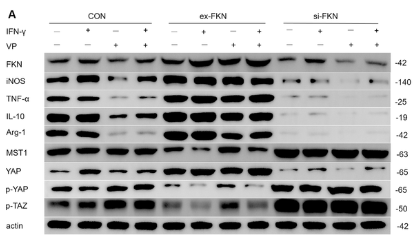
7). FKN regulates macrophage activation in lupus nephritis via the Hippo signaling pathway. Research Square

Application: WB    Species: Mouse    Sample: RAW264.7 cells

Figure 3. FKN regulated polarization in IFN-γ-induced RAW264.7 cells via the Hippo signaling pathway. A, B, C, Western blot analysis and their respective quantitation were performed to assess the expression levels of FKN, MST1, YAP, p-YAP, p-TAZ, iNOS, TNF-α, Arg-1 and IL-10 protein in RAW264.7 cells(full-length blots are presented in Supplementary Figure1). Representative blots are shown. *P

8). 6-paradol molekülünün amiloid beta enjeksiyonu ile oluşturulmuş alzheimer hastalığı modelindeki nöroinflamatuvar ve nöroprotektif etkilerinin araştırılması.

Restrictive clause


Affinity Biosciences tests all products strictly. Citations are provided as a resource for additional applications that have not been validated by Affinity Biosciences. Please choose the appropriate format for each application and consult Materials and Methods sections for additional details about the use of any product in these publications.

For Research Use Only.
Not for use in diagnostic or therapeutic procedures. Not for resale. Not for distribution without written consent. Affinity Biosciences will not be held responsible for patent infringement or other violations that may occur with the use of our products. Affinity Biosciences, Affinity Biosciences Logo and all other trademarks are the property of Affinity Biosciences LTD.