CPT1A Antibody - #DF12004

Related Downloads
Protocols
Product Info
*The optimal dilutions should be determined by the end user.
*Tips:
WB: For western blot detection of denatured protein samples. IHC: For immunohistochemical detection of paraffin sections (IHC-p) or frozen sections (IHC-f) of tissue samples. IF/ICC: For immunofluorescence detection of cell samples. ELISA(peptide): For ELISA detection of antigenic peptide.
Cite Format: Affinity Biosciences Cat# DF12004, RRID:AB_2844809.
Fold/Unfold
Carnitine O palmitoyltransferase 1 liver isoform; Carnitine O palmitoyltransferase I; Carnitine O palmitoyltransferase I liver isoform; Carnitine O-palmitoyltransferase 1; Carnitine O-palmitoyltransferase I; Carnitine palmitoyltransferase 1A (liver); Carnitine palmitoyltransferase 1A; Carnitine palmitoyltransferase I; Carnitine palmitoyltransferase I liver; CPT 1; CPT I; CPT1; CPT1 L; CPT1-L; Cpt1a; CPT1A_HUMAN; CPTI; CPTI-L; L CPT1; liver isoform;
Immunogens
- P50416 CPT1A_HUMAN:
- Protein BLAST With
- NCBI/
- ExPASy/
- Uniprot
MAEAHQAVAFQFTVTPDGIDLRLSHEALRQIYLSGLHSWKKKFIRFKNGIITGVYPASPSSWLIVVVGVMTTMYAKIDPSLGIIAKINRTLETANCMSSQTKNVVSGVLFGTGLWVALIVTMRYSLKVLLSYHGWMFTEHGKMSRATKIWMGMVKIFSGRKPMLYSFQTSLPRLPVPAVKDTVNRYLQSVRPLMKEEDFKRMTALAQDFAVGLGPRLQWYLKLKSWWATNYVSDWWEEYIYLRGRGPLMVNSNYYAMDLLYILPTHIQAARAGNAIHAILLYRRKLDREEIKPIRLLGSTIPLCSAQWERMFNTSRIPGEETDTIQHMRDSKHIVVYHRGRYFKVWLYHDGRLLKPREMEQQMQRILDNTSEPQPGEARLAALTAGDRVPWARCRQAYFGRGKNKQSLDAVEKAAFFVTLDETEEGYRSEDPDTSMDSYAKSLLHGRCYDRWFDKSFTFVVFKNGKMGLNAEHSWADAPIVAHLWEYVMSIDSLQLGYAEDGHCKGDINPNIPYPTRLQWDIPGECQEVIETSLNTANLLANDVDFHSFPFVAFGKGIIKKCRTSPDAFVQLALQLAHYKDMGKFCLTYEASMTRLFREGRTETVRSCTTESCDFVRAMVDPAQTVEQRLKLFKLASEKHQHMYRLAMTGSGIDRHLFCLYVVSKYLAVESPFLKEVLSEPWRLSTSQTPQQQVELFDLENNPEYVSSGGGFGPVADDGYGVSYILVGENLINFHISSKFSCPETDSHRFGRHLKEAMTDIITLFGLSSNSKK
Predictions
Score>80(red) has high confidence and is suggested to be used for WB detection. *The prediction model is mainly based on the alignment of immunogen sequences, the results are for reference only, not as the basis of quality assurance.
High(score>80) Medium(80>score>50) Low(score<50) No confidence
PTMs - P50416 As Substrate
| Site | PTM Type | Enzyme | Source |
|---|---|---|---|
| K86 | Ubiquitination | Uniprot | |
| S106 | Phosphorylation | Uniprot | |
| S158 | Phosphorylation | Uniprot | |
| K161 | Ubiquitination | Uniprot | |
| Y165 | Phosphorylation | Uniprot | |
| K180 | Ubiquitination | Uniprot | |
| K195 | Ubiquitination | Uniprot | |
| K200 | Ubiquitination | Uniprot | |
| K292 | Ubiquitination | Uniprot | |
| T322 | Phosphorylation | Uniprot | |
| S371 | Phosphorylation | Uniprot | |
| K405 | Ubiquitination | Uniprot | |
| K441 | Ubiquitination | Uniprot | |
| Y514 | Phosphorylation | Uniprot | |
| K634 | Ubiquitination | Uniprot | |
| S671 | Phosphorylation | Uniprot | |
| K675 | Ubiquitination | Uniprot | |
| K755 | Ubiquitination | Uniprot | |
| S771 | Phosphorylation | Uniprot | |
| K772 | Ubiquitination | Uniprot |
Research Backgrounds
Catalyzes the transfer of the acyl group of long-chain fatty acid-CoA conjugates onto carnitine, an essential step for the mitochondrial uptake of long-chain fatty acids and their subsequent beta-oxidation in the mitochondrion. Plays an important role in triglyceride metabolism.
Mitochondrion outer membrane>Multi-pass membrane protein.
Strong expression in kidney and heart, and lower in liver and skeletal muscle.
Homohexamer and homotrimer. Identified in a complex that contains at least CPT1A, ACSL1 and VDAC1. Also identified in complexes with ACSL1 and VDAC2 and VDAC3 (By similarity).
A conformation change in the N-terminal region spanning the first 42 residues plays an important role in the regulation of enzyme activity by malonyl-CoA.
Belongs to the carnitine/choline acetyltransferase family.
Research Fields
· Environmental Information Processing > Signal transduction > AMPK signaling pathway. (View pathway)
· Human Diseases > Endocrine and metabolic diseases > Insulin resistance.
· Metabolism > Lipid metabolism > Fatty acid degradation.
· Metabolism > Global and overview maps > Fatty acid metabolism.
· Organismal Systems > Endocrine system > PPAR signaling pathway.
· Organismal Systems > Endocrine system > Adipocytokine signaling pathway.
· Organismal Systems > Endocrine system > Glucagon signaling pathway.
References
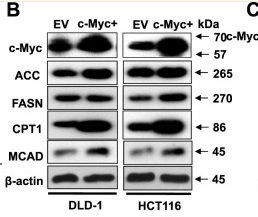
Application: WB Species: Mouse Sample: xenograft tissues
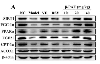
Application: WB Species: Human Sample: L02 cell
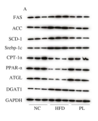
Application: WB Species: Mouse Sample:
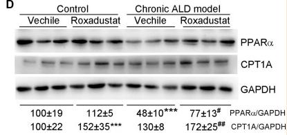
Application: WB Species: Human Sample: HepG2 cells
Application: WB Species: mouse Sample: livers
Restrictive clause
Affinity Biosciences tests all products strictly. Citations are provided as a resource for additional applications that have not been validated by Affinity Biosciences. Please choose the appropriate format for each application and consult Materials and Methods sections for additional details about the use of any product in these publications.
For Research Use Only.
Not for use in diagnostic or therapeutic procedures. Not for resale. Not for distribution without written consent. Affinity Biosciences will not be held responsible for patent infringement or other violations that may occur with the use of our products. Affinity Biosciences, Affinity Biosciences Logo and all other trademarks are the property of Affinity Biosciences LTD.