Product: MyoD1 Antibody
Catalog: AF7733
Description: Rabbit polyclonal antibody to MyoD1
Application: WB IHC IF/ICC
Reactivity: Human, Mouse, Rat, Sheep, Monkey
Prediction: Pig, Zebrafish, Bovine, Sheep, Rabbit, Dog, Chicken, Xenopus
Mol.Wt.: 40~55kDa; 35kD(Calculated).
Uniprot: P15172
RRID: AB_2844097

View similar products>>

   Size Price Inventory
 50ul $250 In stock
 100ul $350 In stock
 200ul $450 In stock

Lead Time: Same day delivery

For pricing and ordering contact:
Local distributors

Product Info

Source:
Rabbit
Application:
WB 1:500-1:2000, IHC 1:50-1:200, IF/ICC 1:100-1:500
*The optimal dilutions should be determined by the end user.
*Tips:

WB: For western blot detection of denatured protein samples. IHC: For immunohistochemical detection of paraffin sections (IHC-p) or frozen sections (IHC-f) of tissue samples. IF/ICC: For immunofluorescence detection of cell samples. ELISA(peptide): For ELISA detection of antigenic peptide.

Reactivity:
Human,Mouse,Rat,Sheep,Monkey
Prediction:
Pig(100%), Zebrafish(100%), Bovine(100%), Rabbit(100%), Dog(100%), Chicken(100%), Xenopus(100%)
Clonality:
Polyclonal
Specificity:
MyoD1 Antibody detects endogenous levels of total MyoD1.
RRID:
AB_2844097
Cite Format: Affinity Biosciences Cat# AF7733, RRID:AB_2844097.
Conjugate:
Unconjugated.
Purification:
The antiserum was purified by peptide affinity chromatography using SulfoLink™ Coupling Resin (Thermo Fisher Scientific).
Storage:
Rabbit IgG in phosphate buffered saline , pH 7.4, 150mM NaCl, 0.02% sodium azide and 50% glycerol. Store at -20 °C. Stable for 12 months from date of receipt.
Alias:

Fold/Unfold

bHLHc1; Class C basic helix-loop-helix protein 1; MYF 3; Myf-3; MYF3; Myoblast determination protein 1; Myod 1; MYOD; MYOD1; MYOD1_HUMAN; Myogenic differentiation 1; Myogenic factor 3; Myogenic factor MYF 3; Myogenin D1; PUM;

Immunogens

Immunogen:
Uniprot:
Gene(ID):
Sequence:
MELLSPPLRDVDLTAPDGSLCSFATTDDFYDDPCFDSPDLRFFEDLDPRLMHVGALLKPEEHSHFPAAVHPAPGAREDEHVRAPSGHHQAGRCLLWACKACKRKTTNADRRKAATMRERRRLSKVNEAFETLKRCTSSNPNQRLPKVEILRNAIRYIEGLQALLRDQDAAPPGAAAAFYAPGPLPPGRGGEHYSGDSDASSPRSNCSDGMMDYSGPPSGARRRNCYEGAYYNEAPSEPRPGKSAAVSSLDCLSSIVERISTESPAAPALLLADVPSESPPRRQEAAAPSEGESSGDPTQSPDAAPQCPAGANPNPIYQVL

Predictions

Predictions:

Score>80(red) has high confidence and is suggested to be used for WB detection. *The prediction model is mainly based on the alignment of immunogen sequences, the results are for reference only, not as the basis of quality assurance.

Species
Results
Score
Pig
100
Bovine
100
Sheep
100
Dog
100
Xenopus
100
Zebrafish
100
Chicken
100
Rabbit
100
Horse
0
Model Confidence:
High(score>80) Medium(80>score>50) Low(score<50) No confidence

PTMs - P15172 As Substrate

Site PTM Type Enzyme
S5 Phosphorylation P06493 (CDK1)
Y30 Phosphorylation P00519 (ABL1)
K58 Acetylation
K99 Acetylation
K102 Acetylation
K104 Acetylation
K104 Methylation
T115 Phosphorylation P17252 (PRKCA)
Y156 Phosphorylation Q02750 (MAP2K1)
S200 Phosphorylation P53778 (MAPK12) , P06493 (CDK1) , P24941 (CDK2)
S201 Phosphorylation P53778 (MAPK12)
S204 Phosphorylation
S207 Phosphorylation
Y213 Phosphorylation
S214 Phosphorylation

Research Backgrounds

Function:

Acts as a transcriptional activator that promotes transcription of muscle-specific target genes and plays a role in muscle differentiation. Together with MYF5 and MYOG, co-occupies muscle-specific gene promoter core region during myogenesis. Induces fibroblasts to differentiate into myoblasts. Interacts with and is inhibited by the twist protein. This interaction probably involves the basic domains of both proteins (By similarity).

PTMs:

Phosphorylated by CDK9. This phosphorylation promotes its function in muscle differentiation.

Acetylated by a complex containing EP300 and PCAF. The acetylation is essential to activate target genes. Conversely, its deacetylation by SIRT1 inhibits its function (By similarity).

Ubiquitinated on the N-terminus; which is required for proteasomal degradation.

Methylation at Lys-104 by EHMT2/G9a inhibits myogenic activity.

Subcellular Location:

Nucleus.

Extracellular region or secreted Cytosol Plasma membrane Cytoskeleton Lysosome Endosome Peroxisome ER Golgi apparatus Nucleus Mitochondrion Manual annotation Automatic computational assertionSubcellular location
Subunit Structure:

Efficient DNA binding requires dimerization with another bHLH protein. Seems to form active heterodimers with ITF-2. Interacts with SUV39H1. Interacts with DDX5. Interacts with CHD2. Interacts with TSC22D3 (By similarity). Interacts with SETD3 (By similarity). Interacts with P-TEFB complex; promotes the transcriptional activity of MYOD1 through its CDK9-mediated phosphorylation (By similarity). Interacts with CSRP3. Interacts with NUPR1 (By similarity).

References

1). Sympathetic activity is correlated with satellite cell aging and myogenesis via β2-adrenoceptor. Stem Cell Research & Therapy (PubMed: 34530910) [IF=7.5]

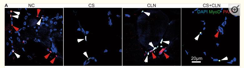
Application: IF/ICC    Species: Mice    Sample: SC cells

Fig. 5 CLB promoted skeletal muscle repair by affecting MyoD and Myf5. A–C CS decreased the number of PAX7+ cells (SCs), but the ratio of MyoD+/MyoD+PAX7+ cells was increased significantly. That is, CS led to the activation of SCs, which hindered their ability to maintain their stemness. CS + CLB treatment prevented this trend. In addition, the PAX7+ cell level and the ratio of MyoD+/MyoD+ PAX7+ cells were not significantly changed by CLB alone in normal mice (F = 15.363 and 5.911, P < 0.001 and = 0.03). D, E On the 10th and 42nd days after injury, the number of Myf5− PAX7+ cells (SCs) in the CS + CLB + injury group was significantly increased compared with that in the CS + injury group (F = 51.219, P < 0.001). F–I On the 10th and 42nd days after injury, the CS + injury group had the lowest number of myonuclei per muscle fiber (transverse section), and this effect was rescued by CLB. The CS + injury group had the largest fibrotic area (F = 11.644, 16.573, both P < 0.001) and the smallest muscle area (F = 11.644, 16.573, both P < 0.001), and this effect was rescued by CLB on both the 10th and 42nd days after injury. One-way ANOVA and Tukey’s test, ns, not significant, # all P < 0.05 compared with all other groups *P < 0.05; **P < 0.001

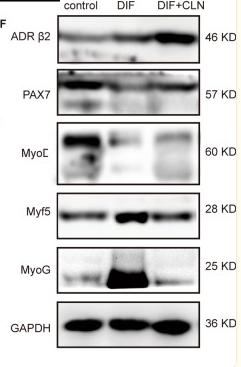
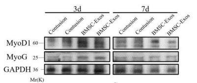
Application: WB    Species: Mice    Sample: SC cells

Fig. 6 CLB-inhibited myogenic differentiation. A, B After 5 days of differentiation, numerous fused myotubes were observed, while CLB (10 μm, 100 μm) significantly inhibited myotube formation (F = 28.9, P < 0.001). C–E After 48 h of myogenic differentiation, the levels of MyoD+ nuclei and β2-ADR were significantly increased, while CLB significantly decreased the expression of MyoD and further increased the expression of β2-ADR (F = 9.642, 8.157, both P < 0.001). F, G After 48 h of differentiation, β2-ADR expression was increased, and CLB further promoted this increase (F = 115.712, P < 0.001). PAX7 expression was decreased after differentiation, and CLB reversed this trend (F = 18.155, P < 0.001). MyoD, Myf5, and MyoG expressions were increased significantly after differentiation, and CLB significantly mitigated these increments (F = 16.324, 8.647, and 154.647, all P < 0.001). All differences were determined using one-way ANOVA and Tukey’s test. ns, not significant, *P < 0.01; **P < 0.001

2). Calycosin inhibited autophagy and oxidative stress in chronic kidney disease skeletal muscle atrophy by regulating AMPK/SKP2/CARM1 signalling pathway. JOURNAL OF CELLULAR AND MOLECULAR MEDICINE (PubMed: 32910538) [IF=5.3]

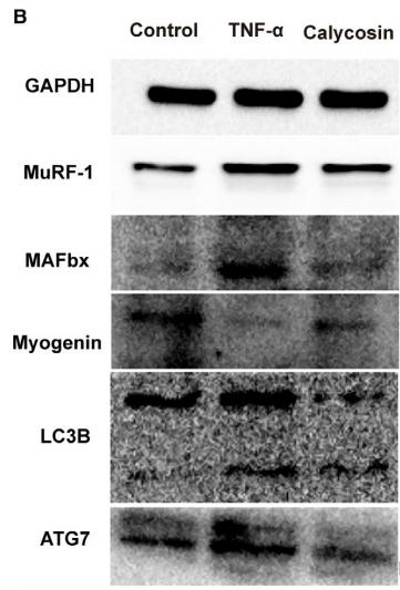
Application: WB    Species: mouse    Sample: C2C12 cells

FIGURE 8| Effect of calycosin on the expression of proteins associated with the AMPK/SKP2/CARM1 signalling pathway in TNF-αinduced C2C12 cells in vitro. A, Representative images of H3R17me2a in C2C12 cells treated with TNF-α or calycosin. B-C, Representative immunoblot of key proteins associated with the AMPK/SKP2/CARM1 signalling pathway in C2C12 cells treated with TNF-α or calycosin.

3). Exosomes derived from inflammatory myoblasts promote M1 polarization and break the balance of myoblast proliferation/differentiation. World Journal of Stem Cells (PubMed: 34909122) [IF=4.1]

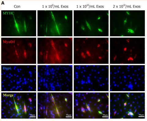
Application: IF/ICC    Species: Human    Sample: macrophages

Figure 7 IF-C2C12-Exos impaired early C2C12 muscle differentiation in vitro. A: Immunofluorescence was used to detect relative expression and distribution of MYHC and Myod1 on day 2 after different treatments. Green, red, and blue signals represent MYHC, Myod1, and nucleus, respectively. Scale bar = 90 μm; B, D and E: Quantification of myotube length, diameter, and fusion rate. Data are presented as mean ± SD. n = 12 or 3. aP < 0.05; C: Representative images of myotube after culture with different concentrations of IF-C2C12-Exosomes for 24 h by Giemsa staining (2 d 2% horse-serum incubation). Scale bar = 100 μm.

4). Bone Marrow Stromal Cell-Derived Exosomes promote muscle healing following contusion through macrophage polarization. STEM CELLS AND DEVELOPMENT (PubMed: 33323007) [IF=4.0]

Application: WB    Species: Mice    Sample:

Figure 5:BMSC-Exos therapy increased the expression of muscle regeneration maker. (A-C) Myod1 and MyoG protein levels in tibialis anterior muscles of contusion and BMSCExos group at 3d and 7d were determined by western blot (n=4). Data are expressed as the means ± standard error of the mean. *P < 0.05. NS: not significant (D) Immunofluorescence was used to detect relative expression and distribution of Pax7 and Myod1 on day 3. Green, red, and blue signal represents Myod1, Pax7, and nucleus, respectively. 200× magnification. Arrows pointed to Pax7 and Myod1 positive cells.

Restrictive clause


Affinity Biosciences tests all products strictly. Citations are provided as a resource for additional applications that have not been validated by Affinity Biosciences. Please choose the appropriate format for each application and consult Materials and Methods sections for additional details about the use of any product in these publications.

For Research Use Only.
Not for use in diagnostic or therapeutic procedures. Not for resale. Not for distribution without written consent. Affinity Biosciences will not be held responsible for patent infringement or other violations that may occur with the use of our products. Affinity Biosciences, Affinity Biosciences Logo and all other trademarks are the property of Affinity Biosciences LTD.