Product: Osteopontin Antibody
Catalog: AF0227
Description: Rabbit polyclonal antibody to Osteopontin
Application: WB IHC IF/ICC
Reactivity: Human, Mouse, Rat
Prediction: Bovine, Dog
Mol.Wt.: 60kDa; 35kD(Calculated).
Uniprot: P10451
RRID: AB_2833402

View similar products>>

   Size Price Inventory
 100ul $280 In stock
 200ul $350 In stock

Lead Time: Same day delivery

For pricing and ordering contact:
Local distributors

Product Info

Source:
Rabbit
Application:
WB 1:500-1:3000, IF/ICC 1:100-1:500, IHC 1:50-1:200
*The optimal dilutions should be determined by the end user.
*Tips:

WB: For western blot detection of denatured protein samples. IHC: For immunohistochemical detection of paraffin sections (IHC-p) or frozen sections (IHC-f) of tissue samples. IF/ICC: For immunofluorescence detection of cell samples. ELISA(peptide): For ELISA detection of antigenic peptide.

Reactivity:
Human,Mouse,Rat
Prediction:
Bovine(88%), Dog(80%)
Clonality:
Polyclonal
Specificity:
Osteopontin Antibody detects endogenous levels of total Osteopontin.
RRID:
AB_2833402
Cite Format: Affinity Biosciences Cat# AF0227, RRID:AB_2833402.
Conjugate:
Unconjugated.
Purification:
The antiserum was purified by peptide affinity chromatography using SulfoLink™ Coupling Resin (Thermo Fisher Scientific).
Storage:
Rabbit IgG in phosphate buffered saline , pH 7.4, 150mM NaCl, 0.02% sodium azide and 50% glycerol. Store at -20 °C. Stable for 12 months from date of receipt.
Alias:

Fold/Unfold

BNSP; Bone sialoprotein 1; BSP I; BSPI; Early T lymphocyte activation 1; ETA 1; ETA1; MGC110940; Nephropontin; OPN; Osteopontin; osteopontin/immunoglobulin alpha 1 heavy chain constant region fusion protein; OSTP_HUMAN; PSEC0156; secreted phosphoprotein 1 (osteopontin, bone sialoprotein I, early T-lymphocyte activation 1); Secreted phosphoprotein 1; SPP 1; SPP-1; SPP1; SPP1/CALPHA1 fusion; Urinary stone protein; Uropontin;

Immunogens

Immunogen:
Uniprot:
Gene(ID):
Expression:
P10451 OSTP_HUMAN:

Bone. Found in plasma.

Description:
osteopontin Binds tightly to hydroxyapatite. Appears to form an integral part of the mineralized matrix. Probably important to cell-matrix interaction. Belongs to the osteopontin family. Ligand for integrin alpha-V/beta-3. 4 isoforms of the human protein are produced by alternative splicing.
Sequence:
MRIAVICFCLLGITCAIPVKQADSGSSEEKQLYNKYPDAVATWLNPDPSQKQNLLAPQNAVSSEETNDFKQETLPSKSNESHDHMDDMDDEDDDDHVDSQDSIDSNDSDDVDDTDDSHQSDESHHSDESDELVTDFPTDLPATEVFTPVVPTVDTYDGRGDSVVYGLRSKSKKFRRPDIQYPDATDEDITSHMESEELNGAYKAIPVAQDLNAPSDWDSRGKDSYETSQLDDQSAETHSHKQSRLYKRKANDESNEHSDVIDSQELSKVSREFHSHEFHSHEDMLVVDPKSKEEDKHLKFRISHELDSASSEVN

Predictions

Predictions:

Score>80(red) has high confidence and is suggested to be used for WB detection. *The prediction model is mainly based on the alignment of immunogen sequences, the results are for reference only, not as the basis of quality assurance.

Species
Results
Score
Bovine
88
Dog
80
Sheep
78
Pig
73
Horse
67
Rabbit
33
Xenopus
0
Zebrafish
0
Chicken
0
Model Confidence:
High(score>80) Medium(80>score>50) Low(score<50) No confidence

PTMs - P10451 As Substrate

Site PTM Type Enzyme
S24 Phosphorylation
S26 Phosphorylation
S27 Phosphorylation
S49 O-Glycosylation
S49 Phosphorylation
S62 Phosphorylation
S63 Phosphorylation
T66 Phosphorylation
S76 Phosphorylation
S78 Phosphorylation
S81 Phosphorylation
S99 Phosphorylation
S102 Phosphorylation
S105 Phosphorylation
S108 Phosphorylation
S117 Phosphorylation
S120 Phosphorylation
S123 Phosphorylation
S126 Phosphorylation
S129 Phosphorylation
T134 O-Glycosylation
T134 Phosphorylation
T138 O-Glycosylation
T138 Phosphorylation
T143 O-Glycosylation
T143 Phosphorylation
T147 O-Glycosylation
T147 Phosphorylation
T152 O-Glycosylation
T152 Phosphorylation
Y165 Phosphorylation
T185 Phosphorylation
T190 Phosphorylation
S191 Phosphorylation
S195 Phosphorylation
Y202 Phosphorylation
S215 Phosphorylation
S219 Phosphorylation
S224 Phosphorylation
Y225 Phosphorylation
T227 Phosphorylation
S228 Phosphorylation
S234 Phosphorylation
T237 Phosphorylation
S239 Phosphorylation
S243 Phosphorylation
S254 Phosphorylation
S258 Phosphorylation
S263 Phosphorylation
S267 Phosphorylation
S270 Phosphorylation
S275 Phosphorylation
S280 Phosphorylation
S291 Phosphorylation
S303 Phosphorylation
S308 Phosphorylation
S310 Phosphorylation
S311 Phosphorylation

Research Backgrounds

Function:

Binds tightly to hydroxyapatite. Appears to form an integral part of the mineralized matrix. Probably important to cell-matrix interaction.

Acts as a cytokine involved in enhancing production of interferon-gamma and interleukin-12 and reducing production of interleukin-10 and is essential in the pathway that leads to type I immunity.

PTMs:

Extensively phosphorylated by FAM20C in the extracellular medium at multiple sites within the S-x-E/pS motif.

O-glycosylated. Isoform 5 is GalNAc O-glycosylated at Thr-59 or Ser-62.

Subcellular Location:

Secreted.

Extracellular region or secreted Cytosol Plasma membrane Cytoskeleton Lysosome Endosome Peroxisome ER Golgi apparatus Nucleus Mitochondrion Manual annotation Automatic computational assertionSubcellular location
Tissue Specificity:

Bone. Found in plasma.

Subunit Structure:

Ligand for integrin alpha-V/beta-3.

Family&Domains:

Belongs to the osteopontin family.

Research Fields

· Cellular Processes > Cellular community - eukaryotes > Focal adhesion.   (View pathway)

· Environmental Information Processing > Signal transduction > PI3K-Akt signaling pathway.   (View pathway)

· Environmental Information Processing > Signal transduction > Apelin signaling pathway.   (View pathway)

· Environmental Information Processing > Signaling molecules and interaction > ECM-receptor interaction.   (View pathway)

· Human Diseases > Infectious diseases: Viral > Human papillomavirus infection.

· Organismal Systems > Immune system > Toll-like receptor signaling pathway.   (View pathway)

References

1). A Janus-Ros Healing System Promoting Infectious Bone Regeneration via Sono-Epigenetic Modulation. Advanced Materials (PubMed: 37855420) [IF=29.4]

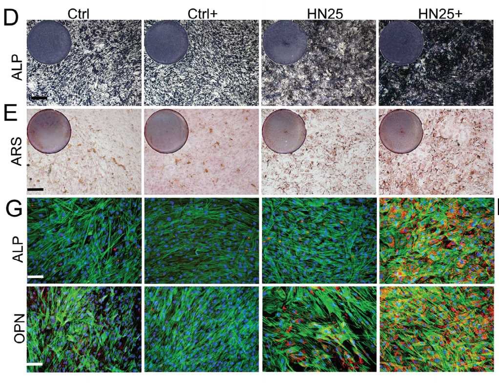
Application: IF/ICC    Species: Rat    Sample: Bone

Figure 4. Biocompatibility and osteogenic differentiation ability of HN25. G) Immunofluorescence staining of ALP and OPN after culturing 14 d on different samples. Scale bar = 100 μm. H) Volcano plot showing differently expressed genes in MSCs from HN25 under US irradiation compared to the control.

2). Self-Adaptive MoO3−x Subnanometric Wires Incorporated Scaffolds for Osteosarcoma Therapy and Bone Regeneration. Advanced Functional Materials [IF=19.0]

3). Osteoblastic and anti-osteoclastic activities of strontium-substituted silicocarnotite ceramics: In vitro and in vivo studies. Bioactive Materials (PubMed: 32280833) [IF=18.9]

4). Fusion peptide engineered “statically-versatile” titanium implant simultaneously enhancing anti-infection, vascularization and osseointegration. Biomaterials (PubMed: 33069134) [IF=14.0]

5). On-demand storage and release of antimicrobial peptides using Pandora's box-like nanotubes gated with a bacterial infection-responsive polymer. Theranostics (PubMed: 31903109) [IF=12.4]

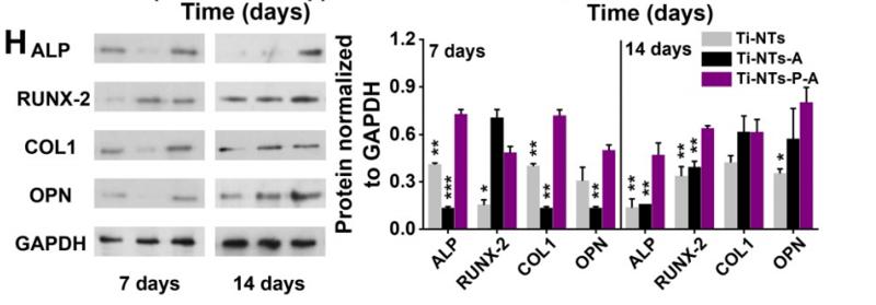
Application: WB    Species: Human    Sample: Human bone mesenchymal stem cells(hBMSCs)

Figure 5. In vitro biocompatibilities and osteogenic activities of the substrates. (A) CCK-8 assay of hBMSCs on the indicated surfaces after 1, 3 and 7 days of culture. * denotes p < 0.05 and ** denotes p < 0.01 compared to Ti-NTs, # denotes p < 0.05 compared to the corresponding control group without peptides. Error bars denote the standard deviations over quadruplicate measurements with separately implants. (B) Confocal fluorescence microscopy images of hBMSCs stained with vinculin, F-actin and DAPI after being cultured for 24 h. Scale bar, 50 μm. (C-F) qRT-PCR assay of osteogenic gene expression of (C) ALP, (D) RUNX-2, (E) COL1 and (F) OPN of hBMSCs after 7 and 14 days of culture. * denotes p < 0.05, ** denotes p < 0.01 and *** denotes p < 0.001 compared to Ti-NTs-P-A; # denotes p < 0.05, & denotes p < 0.01 and $ denotes p < 0.001 compared to the corresponding control group without peptides. All error bars denote the standard deviations over quadruplicate measurements with separately implants. (G) Immunofluorescence staining of hBMSCs cultured on Ti-NTs-P-A for 7 days (ALP and RUNX-2) and 14 days (OPN). The images were obtained by confocal fluorescence microscopy. Scale bar, 50 μm. (H) Western blotting of hBMSCs cultured on the substrates for 7 and 14 days. At each time point, left lane was Ti-NTs, middle lane was Ti-NTs-A and right lane was Ti-NTs-P-A. * denotes p < 0.05, ** denotes p < 0.01 and *** denotes p < 0.001 compared to Ti-NTs-P-A. Error bars denote the standard deviations over triplicate measurements with separately Western blotting results.

6). Biomimetic hydroxyapatite coating on the 3D-printed bioactive porous composite ceramic scaffolds promoted osteogenic differentiation via PI3K/AKT/mTOR signaling pathways and facilitated bone regeneration in vivo. Journal of Materials Science & Technology [IF=10.9]

7). Spontaneous formation of MXene-oxidized sono/chemo-dynamic sonosensitizer/nanocatalyst for antibacteria and bone-tissue regeneration. Journal of Nanobiotechnology (PubMed: 37316836) [IF=10.2]

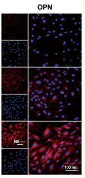
Application: IF/ICC    Species: Rat    Sample: BMMSCs

Fig. 5 In vitro evaluation of C-T@Ti3C2 nanosheets for promoting osteogenesis. a Viability of rat bone marrow mesenchymal stem cells after incubation with different concentrations of C-T@Ti3C2 nanosheets (n = 3 for each group). b CLSM of AM/PI-stained rBMMSCs after various concentrations of C-T@Ti3C2 nanosheets. c CLSM images of the expression of BMP2, OCN, RUNX2 and OPN (at Day 14) in rBMMSCs after different treatments. A representative image of three replicates from each group is shown. d Relative mRNA levels of osteogenic genes (COL-I, BMP2, OPN and RUNX2). e Representative Western blots of total COL-I, OPN, BMP2, RUNX2 and β-actin after induction for 14 days in rBMMSCs. Data are presented as the mean ± SD, and statistical significance was calculated using the two-tailed t test

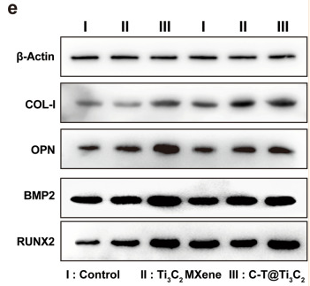
Application: WB    Species: Rat    Sample: BMMSCs

Fig. 5 In vitro evaluation of C-T@Ti3C2 nanosheets for promoting osteogenesis. a Viability of rat bone marrow mesenchymal stem cells after incubation with different concentrations of C-T@Ti3C2 nanosheets (n = 3 for each group). b CLSM of AM/PI-stained rBMMSCs after various concentrations of C-T@Ti3C2 nanosheets. c CLSM images of the expression of BMP2, OCN, RUNX2 and OPN (at Day 14) in rBMMSCs after different treatments. A representative image of three replicates from each group is shown. d Relative mRNA levels of osteogenic genes (COL-I, BMP2, OPN and RUNX2). e Representative Western blots of total COL-I, OPN, BMP2, RUNX2 and β-actin after induction for 14 days in rBMMSCs. Data are presented as the mean ± SD, and statistical significance was calculated using the two-tailed t test

8). A scaffold with zinc-whitlockite nanoparticles accelerates bone reconstruction by promoting bone differentiation and angiogenesis. Nano Research [IF=9.9]

9). Kaempferol alleviates calcium oxalate crystal-induced renal injury and crystal deposition via regulation of the AR/NOX2 signaling pathway. PHYTOMEDICINE (PubMed: 33852977) [IF=7.9]

Application: IHC    Species: Human    Sample: HK-2 cells

Fig. 1. Effect of kaempferol on renal CaOx crystal deposition and tubular injury in vivo. Eligible mice were divided into five groups (n = 8). After a period of 10-day drug treatments, all mice were sacrificed on the 11 th day for the following tests. Pathological results were shown by the (a) images as well as (b-g) corresponding quantitative analysis through HE staining, Pizzolato staining, PAS staining, TUNEL staining, OPN and CD44 immunohistochemistry staining of paraffin embedded kidney sections. (h-j) Renal dysfunction was determined by serum levels of BUN, creatinine and NAGL among the five groups. (k) Group annotations were applied for all histograms. Data were expressed as the fold changes of experimental group to NC group or GA group, and were represented as means ± SD. * p < 0.05 vs. NC group; ** p < 0.05 as GA+Kae (25mg/kg) vs. GA group, GA + Kae (50 mg/kg) vs. GA group, and GA + Kae (25 mg/kg) vs. GA + Kae (50 mg/kg) group, respectively.

10). HA/CD44 Regulates the T Helper 1 Cells Differentiation by Activating Annexin A1/Akt/mTOR Signaling to Drive the Pathogenesis of EAP. Frontiers in Immunology (PubMed: 35693826) [IF=7.3]

Load more

Restrictive clause


Affinity Biosciences tests all products strictly. Citations are provided as a resource for additional applications that have not been validated by Affinity Biosciences. Please choose the appropriate format for each application and consult Materials and Methods sections for additional details about the use of any product in these publications.

For Research Use Only.
Not for use in diagnostic or therapeutic procedures. Not for resale. Not for distribution without written consent. Affinity Biosciences will not be held responsible for patent infringement or other violations that may occur with the use of our products. Affinity Biosciences, Affinity Biosciences Logo and all other trademarks are the property of Affinity Biosciences LTD.