Phospho-TMEM173/STING (Ser366) Antibody - #AF7416
 Antibody. The lane on the left was treated with blocking peptide.")
| Product: | Phospho-TMEM173/STING (Ser366) Antibody |
| Catalog: | AF7416 |
| Description: | Rabbit polyclonal antibody to Phospho-TMEM173/STING (Ser366) |
| Application: | WB IHC IF/ICC |
| Reactivity: | Human, Mouse, Rat |
| Prediction: | Pig, Bovine, Horse, Sheep, Rabbit |
| Mol.Wt.: | 35-40kD; 42kD(Calculated). |
| Uniprot: | Q86WV6 |
| RRID: | AB_2843856 |
Related Downloads
Protocols
Product Info
*The optimal dilutions should be determined by the end user.
*Tips:
WB: For western blot detection of denatured protein samples. IHC: For immunohistochemical detection of paraffin sections (IHC-p) or frozen sections (IHC-f) of tissue samples. IF/ICC: For immunofluorescence detection of cell samples. ELISA(peptide): For ELISA detection of antigenic peptide.
Cite Format: Affinity Biosciences Cat# AF7416, RRID:AB_2843856.
Fold/Unfold
endoplasmic reticulum IFN stimulator; Endoplasmic reticulum interferon stimulator; ERIS; FLJ38577; hMITA; hSTING; Mediator of IRF3 activation; MITA; Mitochondrial mediator of IRF3 activation; MPYS; N terminal methionine proline tyrosine serine plasma membrane tetraspanner; NET23; Stimulator of interferon genes; Stimulator of interferon genes protein; STING; TM173_HUMAN; Tmem173; Transmembrane protein 173;
Immunogens
Ubiquitously expressed. Expressed in skin endothelial cells, alveolar type 2 pneumocytes, bronchial epithelium and alveolar macrophages.
- Q86WV6 STING_HUMAN:
- Protein BLAST With
- NCBI/
- ExPASy/
- Uniprot
MPHSSLHPSIPCPRGHGAQKAALVLLSACLVTLWGLGEPPEHTLRYLVLHLASLQLGLLLNGVCSLAEELRHIHSRYRGSYWRTVRACLGCPLRRGALLLLSIYFYYSLPNAVGPPFTWMLALLGLSQALNILLGLKGLAPAEISAVCEKGNFNVAHGLAWSYYIGYLRLILPELQARIRTYNQHYNNLLRGAVSQRLYILLPLDCGVPDNLSMADPNIRFLDKLPQQTGDHAGIKDRVYSNSIYELLENGQRAGTCVLEYATPLQTLFAMSQYSQAGFSREDRLEQAKLFCRTLEDILADAPESQNNCRLIAYQEPADDSSFSLSQEVLRHLRQEEKEEVTVGSLKTSAVPSTSTMSQEPELLISGMEKPLPLRTDFS
Predictions
Score>80(red) has high confidence and is suggested to be used for WB detection. *The prediction model is mainly based on the alignment of immunogen sequences, the results are for reference only, not as the basis of quality assurance.
High(score>80) Medium(80>score>50) Low(score<50) No confidence
PTMs - Q86WV6 As Substrate
Research Backgrounds
Facilitator of innate immune signaling that acts as a sensor of cytosolic DNA from bacteria and viruses and promotes the production of type I interferon (IFN-alpha and IFN-beta). Innate immune response is triggered in response to non-CpG double-stranded DNA from viruses and bacteria delivered to the cytoplasm. Acts by binding cyclic dinucleotides: recognizes and binds cyclic di-GMP (c-di-GMP), a second messenger produced by bacteria, and cyclic GMP-AMP (cGAMP), a messenger produced by CGAS in response to DNA virus in the cytosol. Upon binding of c-di-GMP or cGAMP, STING1 oligomerizes, translocates from the endoplasmic reticulum and is phosphorylated by TBK1 on the pLxIS motif, leading to recruitment and subsequent activation of the transcription factor IRF3 to induce expression of type I interferon and exert a potent anti-viral state. In addition to promote the production of type I interferons, plays a direct role in autophagy. Following cGAMP-binding, STING1 buds from the endoplasmic reticulum into COPII vesicles, which then form the endoplasmic reticulum-Golgi intermediate compartment (ERGIC). The ERGIC serves as the membrane source for WIPI2 recruitment and LC3 lipidation, leading to formation of autophagosomes that target cytosolic DNA or DNA viruses for degradation by the lysosome. The autophagy- and interferon-inducing activities can be uncoupled and autophagy induction is independent of TBK1 phosphorylation. Autophagy is also triggered upon infection by bacteria: following c-di-GMP-binding, which is produced by live Gram-positive bacteria, promotes reticulophagy (By similarity). Exhibits 2',3' phosphodiester linkage-specific ligand recognition: can bind both 2'-3' linked cGAMP (2'-3'-cGAMP) and 3'-3' linked cGAMP but is preferentially activated by 2'-3' linked cGAMP. The preference for 2'-3'-cGAMP, compared to other linkage isomers is probably due to the ligand itself, whichs adopts an organized free-ligand conformation that resembles the STING1-bound conformation and pays low energy costs in changing into the active conformation. May be involved in translocon function, the translocon possibly being able to influence the induction of type I interferons. May be involved in transduction of apoptotic signals via its association with the major histocompatibility complex class II (MHC-II) (By similarity).
(Microbial infection) Antiviral activity is antagonized by oncoproteins, such as papillomavirus (HPV) protein E7 and adenovirus early E1A protein. Such oncoproteins prevent the ability to sense cytosolic DNA.
Phosphorylation by TBK1 leads to activation and production of IFN-beta. Following cyclic nucleotide (c-di-GMP or cGAMP)-binding, activation and translocation from the endoplasmic reticulum, STING1 is phosphorylated by TBK1 at Ser-366 in the pLxIS motif. The phosphorylated pLxIS motif constitutes an IRF3-binding motif, leading to recruitment of the transcription factor IRF3 to induce type-I interferons and other cytokines. Phosphorylated on tyrosine residues upon MHC-II aggregation (By similarity).
Ubiquitinated. Ubiquitinated via 'Lys-63'-linked ubiquitin chains in response to double-stranded DNA treatment, leading to relocalization to autophagosomes and subsequent degradation; this process is dependent on SQSTM1 (By similarity). 'Lys-63'-linked ubiquitination mediated by TRIM56 at Lys-150 promotes homodimerization and recruitment of the antiviral kinase TBK1 and subsequent production of IFN-beta. 'Lys-48'-linked polyubiquitination at Lys-150 occurring after viral infection is mediated by RNF5 and leads to proteasomal degradation. 'Lys-11'-linked polyubiquitination at Lys-150 by RNF26 leads to stabilize STING1: it protects STING1 from RNF5-mediated 'Lys-48'-linked polyubiquitination.
Endoplasmic reticulum membrane>Multi-pass membrane protein. Cytoplasm>Perinuclear region. Endoplasmic reticulum-Golgi intermediate compartment membrane>Multi-pass membrane protein. Cytoplasmic vesicle>Autophagosome membrane>Multi-pass membrane protein. Mitochondrion outer membrane>Multi-pass membrane protein. Cell membrane>Multi-pass membrane protein.
Note: In response to double-stranded DNA stimulation, translocates from the endoplasmic reticulum through the endoplasmic reticulum-Golgi intermediate compartment and Golgi to post-Golgi vesicles, where the kinase TBK1 is recruited (PubMed:19433799, PubMed:30842659, PubMed:30842653, PubMed:29694889). Upon cGAMP-binding, translocates to the endoplasmic reticulum-Golgi intermediate compartment (ERGIC) in a process that is dependent on COPII vesicles; STING1-containing ERGIC serves as a membrane source for LC3 lipidation, which is a key step in autophagosome biogenesis (PubMed:30842662).
Ubiquitously expressed. Expressed in skin endothelial cells, alveolar type 2 pneumocytes, bronchial epithelium and alveolar macrophages.
Homodimer; forms a homodimer in absence of cyclic nucleotide (c-di-GMP or cGAMP); 'Lys-63'-linked ubiquitination at Lys-150 is required for homodimerization. Homotetramer; in presence of cyclic nucleotide (c-di-GMP or cGAMP), forms tetramers and higher-order oligomers through side-by-side packing (Probable). Interacts (when phosphorylated) with IRF3; following activation and phosphorylation on the pLxIS motif by TBK1, recruits IRF3. Interacts with DDX58/RIG-I, MAVS and SSR2. Interacts with RNF5 and TRIM56. Interacts with TBK1; when homodimer, leading to subsequent production of IFN-beta. Interacts with IFIT1 and IFIT2. Interacts with TRIM29; this interaction induces STING1 ubiquitination and subsequent degradation. Associates with the MHC-II complex (By similarity). Interacts with SEC24C; promoting translocation to the COPII vesicles. Interacts (when ubiquitinated) with SQSTM1; leading to relocalization to autophagosomes (By similarity). Interacts with SURF4. Interacts with HNRNPA2B1.
(Microbial infection) Interacts with human papillomavirus (HPV) protein E7.
(Microbial infection) Interacts with adenovirus early E1A protein.
(Microbial infection) Interacts with herpes simplex virus 1 protein ICP34.5; this interaction inhibits the intracellular DNA sensing pathway.
In absence of cGAMP, the transmembrane and cytoplasmic regions interact to form an integrated, domain-swapped dimeric assembly (By similarity). In absence of cyclic nucleotide (c-di-GMP or cGAMP), the protein is autoinhibited by an intramolecular interaction between the cyclic dinucleotide-binding domain (CBD) and the C-terminal tail (CTT) (PubMed:22579474, PubMed:22705373, PubMed:22728658, PubMed:22728660, PubMed:22728659). Following cGAMP-binding, the cyclic dinucleotide-binding domain (CBD) is closed, leading to a 180 degrees rotation of the CBD domain relative to the transmembrane domain. This rotation is coupled to a conformational change in a loop on the side of the CBD dimer, which leads to the formation of the STING1 tetramer and higher-order oligomers through side-by-side packing (By similarity). The N-terminal part of the CBD region was initially though to contain a fifth transmembrane region (TM5) but is part of the folded, soluble CBD (PubMed:22579474, PubMed:22705373, PubMed:22728658, PubMed:22728660, PubMed:22728659).
The pLxIS motif constitutes an IRF3-binding motif: following phosphorylation by TBK1, the phosphorylated pLxIS motif of STING1 recruits IRF3 (PubMed:25636800). IRF3 is then phosphorylated and activated by TBK1 to induce type-I interferons and other cytokines (PubMed:25636800).
Belongs to the STING family.
Research Fields
· Organismal Systems > Immune system > NOD-like receptor signaling pathway. (View pathway)
· Organismal Systems > Immune system > RIG-I-like receptor signaling pathway. (View pathway)
· Organismal Systems > Immune system > Cytosolic DNA-sensing pathway. (View pathway)
References
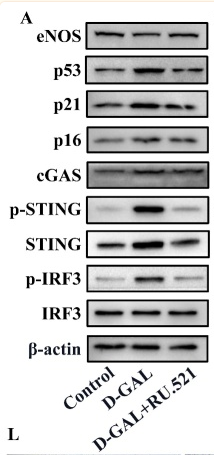
Application: WB Species: Mouse Sample:
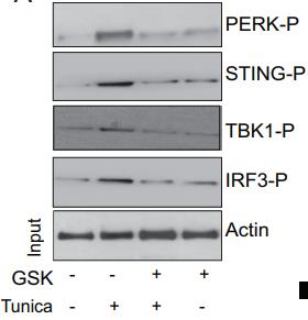
Application: WB Species: mouse Sample: primary cortical neuronal lysate
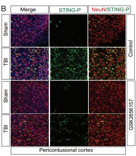
Application: IF/ICC Species: mouse Sample: neurons
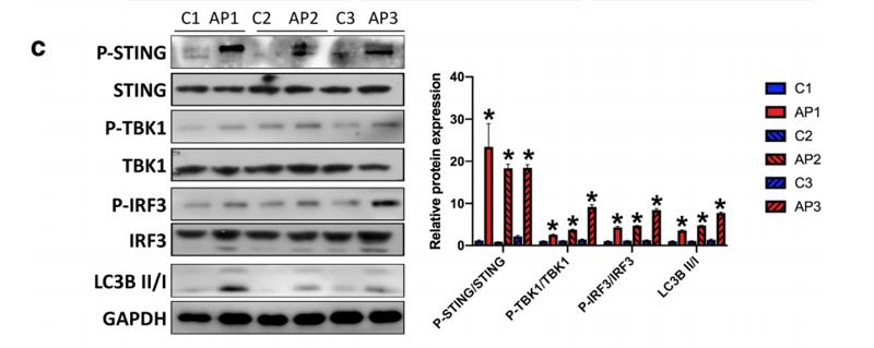
Application: WB Species: rat Sample: pancreatic acinar cells
Restrictive clause
Affinity Biosciences tests all products strictly. Citations are provided as a resource for additional applications that have not been validated by Affinity Biosciences. Please choose the appropriate format for each application and consult Materials and Methods sections for additional details about the use of any product in these publications.
For Research Use Only.
Not for use in diagnostic or therapeutic procedures. Not for resale. Not for distribution without written consent. Affinity Biosciences will not be held responsible for patent infringement or other violations that may occur with the use of our products. Affinity Biosciences, Affinity Biosciences Logo and all other trademarks are the property of Affinity Biosciences LTD.