SLC9A1 Antibody - #DF9933

| Product: | SLC9A1 Antibody |
| Catalog: | DF9933 |
| Description: | Rabbit polyclonal antibody to SLC9A1 |
| Application: | WB IHC IF/ICC |
| Reactivity: | Human, Mouse, Rat, Monkey |
| Prediction: | Pig, Bovine, Horse, Sheep, Rabbit, Dog, Chicken, Xenopus |
| Mol.Wt.: | 91 kDa; 91kD(Calculated). |
| Uniprot: | P19634 |
| RRID: | AB_2843127 |
Related Downloads
Protocols
Product Info
*The optimal dilutions should be determined by the end user.
*Tips:
WB: For western blot detection of denatured protein samples. IHC: For immunohistochemical detection of paraffin sections (IHC-p) or frozen sections (IHC-f) of tissue samples. IF/ICC: For immunofluorescence detection of cell samples. ELISA(peptide): For ELISA detection of antigenic peptide.
Cite Format: Affinity Biosciences Cat# DF9933, RRID:AB_2843127.
Fold/Unfold
amiloride-sensitive; APNH; APNH1; FLJ42224; Na Li countertransporter; Na(+)/H(+) antiporter; Na(+)/H(+) exchanger 1; Na+ H+ antiporter amiloride-sensitive; Na+ H+ antiporter; Na+ H+ exchanger 1; NHE-1; NHE1; OTTHUMP00000004468; SL9A1_HUMAN; SLC9A1; Sodium hydrogen exchanger 1; Sodium/hydrogen exchanger 1; solute carrier family 9; Solute carrier family 9 member 1; Solute carrier family 9 sodium hydrogen exchanger isoform 1 antiporter Na+ H+ amiloride sensitive; Solute carrier family 9 subfamily A (NHE1 cation proton antiporter 1) member 1; Solute carrier family 9 subfamily A member 1;
Immunogens
- P19634 SL9A1_HUMAN:
- Protein BLAST With
- NCBI/
- ExPASy/
- Uniprot
MVLRSGICGLSPHRIFPSLLVVVALVGLLPVLRSHGLQLSPTASTIRSSEPPRERSIGDVTTAPPEVTPESRPVNHSVTDHGMKPRKAFPVLGIDYTHVRTPFEISLWILLACLMKIGFHVIPTISSIVPESCLLIVVGLLVGGLIKGVGETPPFLQSDVFFLFLLPPIILDAGYFLPLRQFTENLGTILIFAVVGTLWNAFFLGGLMYAVCLVGGEQINNIGLLDNLLFGSIISAVDPVAVLAVFEEIHINELLHILVFGESLLNDAVTVVLYHLFEEFANYEHVGIVDIFLGFLSFFVVALGGVLVGVVYGVIAAFTSRFTSHIRVIEPLFVFLYSYMAYLSAELFHLSGIMALIASGVVMRPYVEANISHKSHTTIKYFLKMWSSVSETLIFIFLGVSTVAGSHHWNWTFVISTLLFCLIARVLGVLGLTWFINKFRIVKLTPKDQFIIAYGGLRGAIAFSLGYLLDKKHFPMCDLFLTAIITVIFFTVFVQGMTIRPLVDLLAVKKKQETKRSINEEIHTQFLDHLLTGIEDICGHYGHHHWKDKLNRFNKKYVKKCLIAGERSKEPQLIAFYHKMEMKQAIELVESGGMGKIPSAVSTVSMQNIHPKSLPSERILPALSKDKEEEIRKILRNNLQKTRQRLRSYNRHTLVADPYEEAWNQMLLRRQKARQLEQKINNYLTVPAHKLDSPTMSRARIGSDPLAYEPKEDLPVITIDPASPQSPESVDLVNEELKGKVLGLSRDPAKVAEEDEDDDGGIMMRSKETSSPGTDDVFTPAPSDSPSSQRIQRCLSDPGPHPEPGEGEPFFPKGQ
Predictions
Score>80(red) has high confidence and is suggested to be used for WB detection. *The prediction model is mainly based on the alignment of immunogen sequences, the results are for reference only, not as the basis of quality assurance.
High(score>80) Medium(80>score>50) Low(score<50) No confidence
PTMs - P19634 As Substrate
| Site | PTM Type | Enzyme | Source |
|---|---|---|---|
| S5 | Phosphorylation | Uniprot | |
| S11 | Phosphorylation | Uniprot | |
| S18 | Phosphorylation | Uniprot | |
| T42 | Phosphorylation | Uniprot | |
| N75 | N-Glycosylation | Uniprot | |
| Y96 | Phosphorylation | Uniprot | |
| S158 | Phosphorylation | Uniprot | |
| S372 | Phosphorylation | Uniprot | |
| K559 | Ubiquitination | Uniprot | |
| K560 | Ubiquitination | Uniprot | |
| Y577 | Phosphorylation | Uniprot | |
| K583 | Ubiquitination | Uniprot | |
| S599 | Phosphorylation | Uniprot | |
| S602 | Phosphorylation | Uniprot | |
| T603 | Phosphorylation | Uniprot | |
| S605 | Phosphorylation | Uniprot | |
| K612 | Ubiquitination | Uniprot | |
| S616 | Phosphorylation | Uniprot | |
| S624 | Phosphorylation | Uniprot | |
| K625 | Ubiquitination | Uniprot | |
| S648 | Phosphorylation | P31749 (AKT1) | Uniprot |
| T653 | Phosphorylation | O75116 (ROCK2) , Q13464 (ROCK1) | Uniprot |
| Y659 | Phosphorylation | Uniprot | |
| Y683 | Phosphorylation | Uniprot | |
| T685 | Phosphorylation | Uniprot | |
| S693 | Phosphorylation | P28482 (MAPK1) | Uniprot |
| T695 | Phosphorylation | Uniprot | |
| S697 | Phosphorylation | Uniprot | |
| S703 | Phosphorylation | P31749 (AKT1) , Q15418 (RPS6KA1) | Uniprot |
| Y708 | Phosphorylation | Uniprot | |
| T718 | Phosphorylation | Q16539 (MAPK14) | Uniprot |
| S723 | Phosphorylation | Q16539 (MAPK14) | Uniprot |
| S726 | Phosphorylation | Q16539 (MAPK14) | Uniprot |
| S729 | Phosphorylation | Q16539 (MAPK14) | Uniprot |
| K738 | Ubiquitination | Uniprot | |
| K750 | Ubiquitination | Uniprot | |
| S766 | Phosphorylation | Uniprot | |
| S770 | Phosphorylation | P28482 (MAPK1) | Uniprot |
| S771 | Phosphorylation | Uniprot | |
| T774 | Phosphorylation | Uniprot | |
| T779 | Phosphorylation | Uniprot | |
| S783 | Phosphorylation | Uniprot | |
| S785 | Phosphorylation | P28482 (MAPK1) | Uniprot |
| S787 | Phosphorylation | Uniprot | |
| S788 | Phosphorylation | Uniprot | |
| S796 | Phosphorylation | P31749 (AKT1) | Uniprot |
Research Backgrounds
Involved in pH regulation to eliminate acids generated by active metabolism or to counter adverse environmental conditions. Major proton extruding system driven by the inward sodium ion chemical gradient. Plays an important role in signal transduction.
O-glycosylated.
Ubiquitinated, leading to its degradation by the proteasome. Ubiquitination is reduced by CHP1 (By similarity).
Membrane>Multi-pass membrane protein. Endoplasmic reticulum membrane>Multi-pass membrane protein. Cell membrane>Multi-pass membrane protein.
Note: Colocalizes with CHP1 at the reticulum endoplasmic (By similarity). Colocalizes with CHP1 and CHP2 at the plasma membrane.
Kidney and intestine.
Oligomer (By similarity). Interacts with CALM1 in a calcium-dependent manner. Interacts with TESC. Interacts (via the juxtamembrane region of the cytoplasmic C-terminal domain) with CHP1; the interaction occurs at the plasma membrane in a calcium-dependent manner. Interacts with CHP2; the interaction occurs in a calcium-dependent manner.
Belongs to the monovalent cation:proton antiporter 1 (CPA1) transporter (TC 2.A.36) family.
Research Fields
· Cellular Processes > Cell motility > Regulation of actin cytoskeleton. (View pathway)
· Environmental Information Processing > Signal transduction > cAMP signaling pathway. (View pathway)
· Environmental Information Processing > Signal transduction > Apelin signaling pathway. (View pathway)
· Human Diseases > Cancers: Overview > Proteoglycans in cancer.
· Organismal Systems > Circulatory system > Cardiac muscle contraction. (View pathway)
· Organismal Systems > Circulatory system > Adrenergic signaling in cardiomyocytes. (View pathway)
· Organismal Systems > Endocrine system > Thyroid hormone signaling pathway. (View pathway)
· Organismal Systems > Digestive system > Salivary secretion.
· Organismal Systems > Digestive system > Gastric acid secretion.
· Organismal Systems > Digestive system > Pancreatic secretion.
References

Application: IHC Species: Rat Sample: myocardial tissues
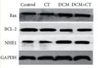
Application: WB Species: Rat Sample: myocardial tissues
Restrictive clause
Affinity Biosciences tests all products strictly. Citations are provided as a resource for additional applications that have not been validated by Affinity Biosciences. Please choose the appropriate format for each application and consult Materials and Methods sections for additional details about the use of any product in these publications.
For Research Use Only.
Not for use in diagnostic or therapeutic procedures. Not for resale. Not for distribution without written consent. Affinity Biosciences will not be held responsible for patent infringement or other violations that may occur with the use of our products. Affinity Biosciences, Affinity Biosciences Logo and all other trademarks are the property of Affinity Biosciences LTD.