MST1 Antibody - #DF8430

| Product: | MST1 Antibody |
| Catalog: | DF8430 |
| Description: | Rabbit polyclonal antibody to MST1 |
| Application: | WB IHC IF/ICC |
| Reactivity: | Human, Mouse, Rat |
| Mol.Wt.: | 85 kDa; 80kD(Calculated). |
| Uniprot: | P26927 |
| RRID: | AB_2841678 |
Product Info
*The optimal dilutions should be determined by the end user.
*Tips:
WB: For western blot detection of denatured protein samples. IHC: For immunohistochemical detection of paraffin sections (IHC-p) or frozen sections (IHC-f) of tissue samples. IF/ICC: For immunofluorescence detection of cell samples. ELISA(peptide): For ELISA detection of antigenic peptide.
Cite Format: Affinity Biosciences Cat# DF8430, RRID:AB_2841678.
Fold/Unfold
D3F15S2; DNF15S2; Hepatocyte growth factor like protein alpha chain; Hepatocyte growth factor like protein; Hepatocyte growth factor like protein beta chain; Hepatocyte growth factor like protein homolog; Hepatocyte growth factor-like protein beta chain; HGFL; HGFL_HUMAN; Macrophage stimulating 1 (hepatocyte growth factor like); Macrophage stimulatory protein; Macrophage-stimulating protein; MSP; MST1; NF15S2; OTTHUMP00000208927;
Immunogens
A synthesized peptide derived from human MST1, corresponding to a region within C-terminal amino acids.
- P26927 HGFL_HUMAN:
- Protein BLAST With
- NCBI/
- ExPASy/
- Uniprot
MGWLPLLLLLTQCLGVPGQRSPLNDFQVLRGTELQHLLHAVVPGPWQEDVADAEECAGRCGPLMDCRAFHYNVSSHGCQLLPWTQHSPHTRLRRSGRCDLFQKKDYVRTCIMNNGVGYRGTMATTVGGLPCQAWSHKFPNDHKYTPTLRNGLEENFCRNPDGDPGGPWCYTTDPAVRFQSCGIKSCREAACVWCNGEEYRGAVDRTESGRECQRWDLQHPHQHPFEPGKFLDQGLDDNYCRNPDGSERPWCYTTDPQIEREFCDLPRCGSEAQPRQEATTVSCFRGKGEGYRGTANTTTAGVPCQRWDAQIPHQHRFTPEKYACKDLRENFCRNPDGSEAPWCFTLRPGMRAAFCYQIRRCTDDVRPQDCYHGAGEQYRGTVSKTRKGVQCQRWSAETPHKPQFTFTSEPHAQLEENFCRNPDGDSHGPWCYTMDPRTPFDYCALRRCADDQPPSILDPPDQVQFEKCGKRVDRLDQRRSKLRVVGGHPGNSPWTVSLRNRQGQHFCGGSLVKEQWILTARQCFSSCHMPLTGYEVWLGTLFQNPQHGEPSLQRVPVAKMVCGPSGSQLVLLKLERSVTLNQRVALICLPPEWYVVPPGTKCEIAGWGETKGTGNDTVLNVALLNVISNQECNIKHRGRVRESEMCTEGLLAPVGACEGDYGGPLACFTHNCWVLEGIIIPNRVCARSRWPAVFTRVSVFVDWIHKVMRLG
PTMs - P26927 As Substrate
| Site | PTM Type | Enzyme | Source |
|---|---|---|---|
| S185 | Phosphorylation | Uniprot | |
| Y199 | Phosphorylation | Uniprot | |
| N296 | N-Glycosylation | Uniprot | |
| S480 | Phosphorylation | Uniprot | |
| K573 | Ubiquitination | Uniprot | |
| N615 | N-Glycosylation | Uniprot |
PTMs - P26927 As Enzyme
Research Backgrounds
Cleaved after Arg-483, probably by HPN/Hepsin, to yield the active form consisting of two disulfide-linked chains.
Secreted.
Dimer of an alpha chain and a beta chain linked by a disulfide bond. Interacts (via beta chain) with MST1R (via SEMA domain).
Belongs to the peptidase S1 family. Plasminogen subfamily.
References
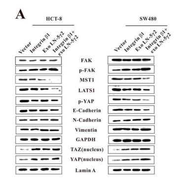
Application: WB Species: Human Sample: HCT-8 and SW480 cells
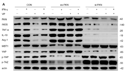
Application: WB Species: Mouse Sample: RAW264.7 cells
Restrictive clause
Affinity Biosciences tests all products strictly. Citations are provided as a resource for additional applications that have not been validated by Affinity Biosciences. Please choose the appropriate format for each application and consult Materials and Methods sections for additional details about the use of any product in these publications.
For Research Use Only.
Not for use in diagnostic or therapeutic procedures. Not for resale. Not for distribution without written consent. Affinity Biosciences will not be held responsible for patent infringement or other violations that may occur with the use of our products. Affinity Biosciences, Affinity Biosciences Logo and all other trademarks are the property of Affinity Biosciences LTD.